. 2021 Mar 1;35(5-6):354-366.
doi: 10.1101/gad.345363.120. Epub 2021 Feb 18.

Polycomb complexes redundantly maintain epidermal stem cell identity during development

Affiliations

Polycomb complexes redundantly maintain epidermal stem cell identity during development

Idan Cohen et al. Genes Dev. .

Abstract

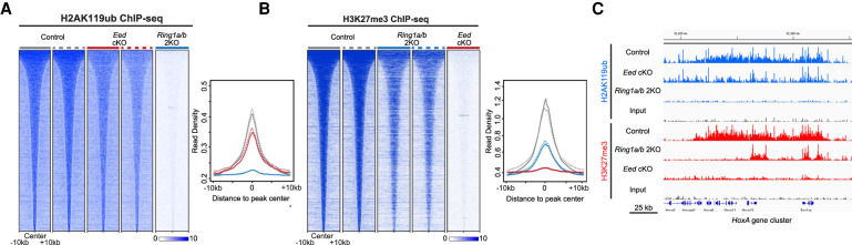
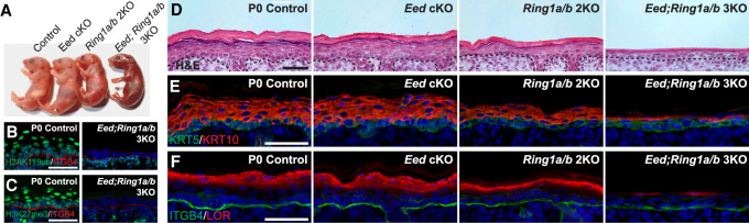
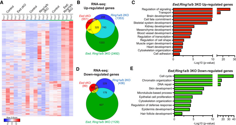
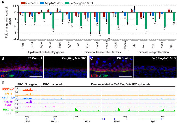
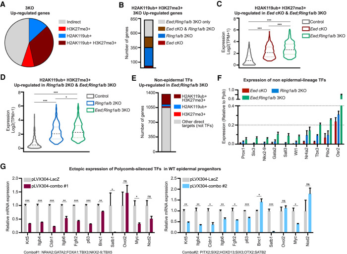
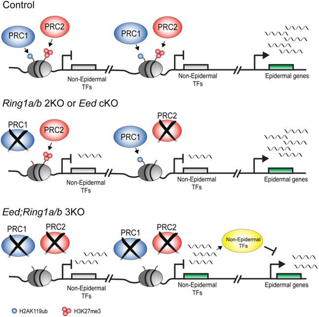
Polycomb repressive complex 1 (PRC1) and PRC2 are critical epigenetic developmental regulators. PRC1 and PRC2 largely overlap in their genomic binding and cooperate to establish repressive chromatin domains demarcated by H2AK119ub and H3K27me3. However, the functional contribution of each complex to gene repression has been a subject of debate, and understanding of its physiological significance requires further studies. Here, using the developing murine epidermis as a paradigm, we uncovered a previously unappreciated functional redundancy between Polycomb complexes. Coablation of PRC1 and PRC2 in embryonic epidermal progenitors resulted in severe defects in epidermal stratification, a phenotype not observed in the single PRC1-null or PRC2-null epidermis. Molecular dissection indicated a loss of epidermal identity that was coupled to a strong derepression of nonlineage transcription factors, otherwise repressed by either PRC1 or PRC2 in the absence of its counterpart. Ectopic expression of subsets of PRC1/2-repressed nonepidermal transcription factors in wild-type epidermal stem cells was sufficient to suppress epidermal identity genes, highlighting the importance of functional redundancy between PRC1 and PRC2. Altogether, our studies show how PRC1 and PRC2 function as two independent counterparts, thereby providing a repressive safety net that protects and preserves lineage identity.

Keywords: H2AK119ub; H3K27me3; PRC1; PRC2; Polycomb; epidermis; epigenetics; skin; stem cell.


Figures

Figure 1.
Figure 1.
Repressive marks of PRC1 and PRC2 are largely maintained in the absence of their counterparts in FACS-purified EpSCs. (A) Read densities of H2AK119ub ChIP-seq peaks in P0 control, Eed cKO, and Ring1a/b 2KO FACS-purified EpSCs, ±10 kb from peak centers. (B) Read densities of H3K27me3 ChIP-seq peaks in P0 control, Ring1a/b 2KO, and Eed cKO FACS-purified EpSCs, ±10 kb from peak centers. (C) Integrative Genomics Viewer (IGV) browser view of ChIP-seq tracks for H2AK119ub and H3K27me3 in P0 control, Eed cKO, and Ring1a/b 2KO FACS-purified EpSCs on the HoxA gene cluster. Y-axis normalized by sequencing depths.
Figure 2.
Figure 2.
Epidermal development is arrested in PRC1/2-null mutants. (A) Gross appearance of newborn (P0) pups for all indicated genotypes. Note that the Eed; Ring1a/b 3KO pup displays red skin, indicating epidermal defects. (B,C) IF staining for H2AK119ub (B; green) and H3K27me3 (C; green) confirming complete loss of these marks in the Eed; Ring1a/b 3KO epidermis. The basement membrane is labeled by Integrin β4 (ITGB4). Scale bar, 50 µm. (D) Hematoxylin and eosin (H&E) analysis of newborn skins. Scale bar, 50 µm. (E) IF staining for basal layer marker KRT5 (green) and suprabasal marker KRT10 (red). Scale bar, 50 µm. (F) IF staining for the late differentiation marker Loricrin (LOR; red). Scale bar, 50 µm.
Figure 3.
Figure 3.
Loss of both PRC1 and PRC2 in EpSCs exacerbates transcriptional changes in single complex mutants. (A) Heat map showing that the DEGs from 3KO were significantly less affected in Eed cKO or Ring1a/b 2KO. The DEGs from 3KO versus control were used. (B) Venn diagram showing overlap of up-regulated genes in P0 Eed cKO, Ring1a/b 2KO, and Ring1a/b; Eed 3KO epidermis. (C) Gene ontology (GO) analysis of up-regulated genes in the Eed; Ring1a/b 3KO epidermis. (D) Venn diagram showing overlap of down-regulated genes in P0 Eed cKO, Ring1a/b 2KO, and Eed; Ring1a/b 3KO epidermis. (E) GO analysis of down-regulated genes in the Eed; Ring1a/b 3KO epidermis.
Figure 4.
Figure 4.
Indirect PRC1 and PRC2 functions preserve EpSC identity. (A) Fold change in epidermal gene expression in P0 Eed cKO, Ring1a/b 2KO, and Eed; Ring1a/b 3KO epidermis. (*) P < 0.05, (**) P < 0.01, (***) P < 0.001. (B,C) IF staining of the epidermal markers p63 (B) and SATB1 (C) in P0 control and Eed; Ring1a/b 3KO epidermis. The basement membrane is labeled by Integrin β4 (ITGB4; green). Scale bars, 50 µm. (D) IGV browser view of Polycomb marks and subunits for PRC1/2 down-regulated genes. Arrows indicate transcription start sites (TSSs) and transcription direction.
Figure 5.
Figure 5.
PRC1 and PRC2 redundantly repress nonlineage transcription factors in EpSCs. (A) Distribution of H3K27me3 and H2AK119ub marks on the up-regulated genes in Eed; Ring1a/b 3KO. (B) Distribution of PRC1 and PRC2 up-regulated direct target genes among P0 Eed cKO, Ring1a/b 2KO, and Eed; Ring1a/b 3KO EpSCs. (C) Expression levels of PRC1 and PRC2 direct target genes in P0 control, Eed cKO, and Eed; Ring1a/b 3KO EpSCs. (D) Expression levels of PRC1 and PRC2 direct target genes in P0 control, Ring1a/b 2KO, and Eed; Ring1a/b 3KO EpSCs. (E) Proportion of up-regulated transcription factor (TF) genes marked by PRC1, PRC2, or PRC1/2. (F) Relative expression levels of nonepidermal TFs in P0 Eed cKO, Ring1a/b 2KO, and Eed; Ring1a/b 3KO EpSCs. (G) Relative expression levels of epidermal genes in keratinocytes ectopically expressing combinations of nonepidermal TFs. Combination 1 included NR4A2, GATA2, FOXA1, TBX3, NKX2-9, and TBX5. Combination 2 included PITX2, SIX2, HOXD13, SIX3, OTX2, and SATB2.
Figure 6.
Figure 6.
Redundant repressive functions of PRC1 and PRC2 promote epidermal cell fate in EpSCs. (Top) In control EpSCs, PRC1 and PRC2 repressive marks are present on the promoters of nonepidermal genes. Epidermal genes are expressed. (Middle) In Eed cKO or Ring1a/b 2KO EpSCs, loss of either PRC complex results in mild derepression of nonepidermal genes. Epidermal gene expression is not suppressed. (Bottom) In Eed; Ring1a/b 3KO EpSCs, loss of both complexes results in a robust derepression of nonepidermal transcription factor (TF) genes, highlighting that PRC1 and PRC2 redundantly repress these genes. Epidermal genes are suppressed in Eed; Ring1a/b 3KO EpSCs, emphasizing the physiological importance of PRC1 and PRC2 functional redundancy in cell identity maintenance.

Similar articles

Cited by

References

    1. Adhikary G, Grun D, Balasubramanian S, Kerr C, Huang JM, Eckert RL. 2015. Survival of skin cancer stem cells requires the Ezh2 polycomb group protein. Carcinogenesis 36: 800–810. 10.1093/carcin/bgv064 - DOI - PMC - PubMed
    1. Allis CD, Jenuwein T. 2016. The molecular hallmarks of epigenetic control. Nat Rev Genet 17: 487–500. 10.1038/nrg.2016.59 - DOI - PubMed
    1. Aloia L, Di Stefano B, Di Croce L. 2013. Polycomb complexes in stem cells and embryonic development. Development 140: 2525–2534. 10.1242/dev.091553 - DOI - PubMed
    1. Atlasi Y, Stunnenberg HG. 2017. The interplay of epigenetic marks during stem cell differentiation and development. Nat Rev Genet 18: 643–658. 10.1038/nrg.2017.57 - DOI - PubMed
    1. Balasubramanian S, Lee K, Adhikary G, Gopalakrishnan R, Rorke EA, Eckert RL. 2008. The Bmi-1 polycomb group gene in skin cancer: regulation of function by (-)-epigallocatechin-3-gallate. Nutr Rev 66: S65–S68. 10.1111/j.1753-4887.2008.00071.x - DOI - PMC - PubMed

Publication types

MeSH terms

Substances