. 2021 Mar 1;35(5-6):329-334.
doi: 10.1101/gad.346460.120. Epub 2021 Feb 18.

Circadian hepatocyte clocks keep synchrony in the absence of a master pacemaker in the suprachiasmatic nucleus or other extrahepatic clocks

Affiliations

Circadian hepatocyte clocks keep synchrony in the absence of a master pacemaker in the suprachiasmatic nucleus or other extrahepatic clocks

Flore Sinturel et al. Genes Dev. .

Abstract

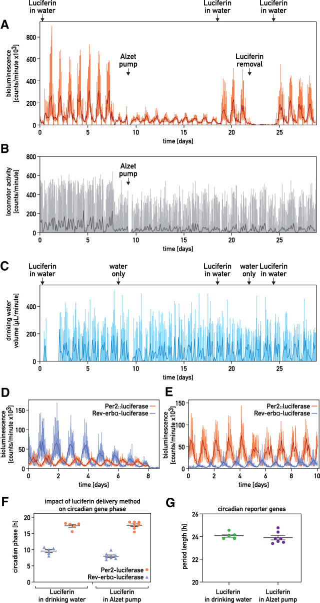
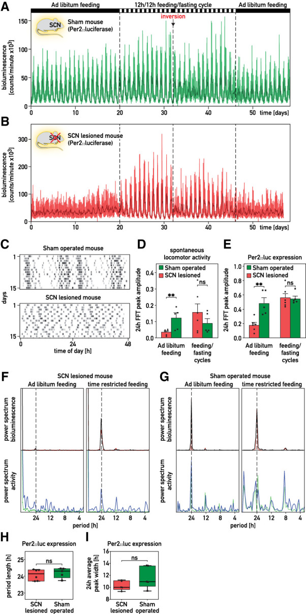
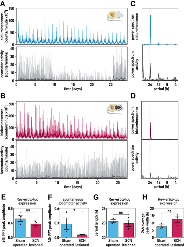
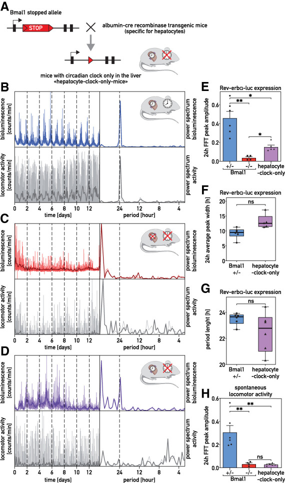
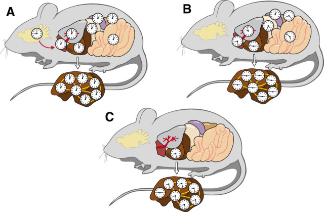
It has been assumed that the suprachiasmatic nucleus (SCN) synchronizes peripheral circadian oscillators. However, this has never been convincingly shown, since biochemical time series experiments are not feasible in behaviorally arrhythmic animals. By using long-term bioluminescence recording in freely moving mice, we show that the SCN is indeed required for maintaining synchrony between organs. Surprisingly, however, circadian oscillations persist in the livers of mice devoid of an SCN or oscillators in cells other than hepatocytes. Hence, similar to SCN neurons, hepatocytes can maintain phase coherence in the absence of Zeitgeber signals produced by other organs or environmental cycles.

Keywords: circadian gene expression; in vivo bioluminescence recording; liver; suprachiasmatic nucleus.


Figures

Figure 1.
Figure 1.
Long-term recording of circadian whole-body bioluminescence. (AC) Whole-body recording of a hairless Per2::luc mouse entrained by a skeleton photoperiod. Luciferin was delivered as indicated. (A) Bioluminescence. (B) Spontaneous locomotor activity. (C) Drinking activity. (Orange, gray, and light blue lines in A–C) Raw data (1-min photon counts, 1-min activity counts, and 1-min water consumption, respectively), (darker lines in AC) 120-min time-binned traces. (D,E) Whole-body Per2::luciferase and hepatic Rev-erbα-luciferase recordings from two different animals kept in constant darkness and receiving luciferin through Alzet pumps (D) or with the drinking water (E). (Light lines) Raw data, (darker lines) 120-min time-binned traces. (F,G) Relationship between the phases (F) and the period lengths (G) of Per2::luciferase and hepatic Rev-erbα-luciferase expression of mice receiving luciferin in the drinking water (n = 5) or by an Alzet micro-osmotic pumps (n = 7). Mice were kept in constant darkness.
Figure 2.
Figure 2.
The SCN is required for the synchronization of peripheral clocks among organs. (A,B) Whole-body bioluminescence for sham-operated (A) and SCN-lesioned (B) Per2::luc mice kept in constant darkness. The feeding regimens are reported above the graphs. The spontaneous locomotor activities of the animals are shown in Supplemental Figure S2A,B. (Thin lines) Raw data (1 min photon counts), (bold lines) 120-min time-binned traces. (C) Actograms of spontaneous locomotor activity of the sham-operated mouse used in A and the SCN-lesioned mouse used in B. After surgery, mice were housed in 12-h/12-h light/dark (LD) cycles and then recorded during 15 d in constant darkness (D/D). (D,E) Amplitude of the FFT peak in the circadian range (≈24 h) of locomotor activities (D) and of Per2::luciferase expression (E) of SCN-lesioned and sham-operated mice subjected to different feeding regimens (means ± SEM, n ≥ 5 per group). (F,G) Periodograms (FFT analysis) of bioluminescence and locomotor activity recorded for the sham-operated mouse displayed in A and the SCN-lesioned mouse displayed in B. (Red and green lines) Raw data (1-min counts), (black and blue lines) integrated data over 120 points. (H,I) Period length from FFT analysis (H) and average oscillation width (I) in sham-operated and SCN-lesioned animals subjected to feeding–fasting cycles. Whiskers of the box plots range from the minimum to the maximum values. The median values are indicated by the horizontal segment within the box, n ≥ 5 per group. Statistical tests for D, E, H, and I were two-tailed Mann–Whitney test. (**) P < 0.01, (ns) nonsignificant.
Figure 3.
Figure 3.
Hepatocyte oscillators maintain synchrony in SCN-lesioned animals. (A,B) Recordings of circadian Rev-erbα-luciferase expression in the livers of sham-operated (blue; A) and SCN-lesioned (pink; B) mice receiving luciferin in the drinking water. Mice transduced with the Adv-Rev-erbα-luc vector were shaved in a dorsal region over the liver. Forty-eight hours after transduction, bioluminescence was monitored in constant darkness (food and water ad libitum). Spontaneous locomotor activity profiles monitored simultaneously are shown below the bioluminescence tracings. The drop of the signal for locomotor activity in the middle of the recording was caused by the obstruction of the IR detector by litter piled up by the mouse. (Thin colored and gray lines ) Raw data (1-min counts), (bold lines) 120-min time-binned traces. (C,D) Periodograms (FFT analysis) of the data shown in A and B. (Thin colored and gray lines) Raw data (1-min counts), (bold lines) 120-min time-binned traces. (E,F) Amplitudes of the FFT peaks in the circadian range of hepatic Rev-erbα-luciferase expression (E) and spontaneous locomotor activity rhythms (F) of SCN-lesioned and sham-operated mice maintained in constant conditions (means ± SEM, n = 3 per group). The statistical test was a one-tailed Mann–Whitney test. (*) P < 0.05, (ns) nonsignificant. (G,H) Period lengths of Rev-erbα-luciferase expression (G) and average oscillation width in the circadian range (≈24 h) (H) of sham-operated and SCN-lesioned animals maintained in constant conditions (means ± SEM, n = 3 per group). The statistical test was a two-tailed Mann–Whitney test. (ns) Nonsignificant.
Figure 4.
Figure 4.
The livers of “hepatocyte clock-only mice” keep partial phase coherence. (A) Scheme of the lox-stop allele used to generate “hepatocyte clock-only mice.” (BD) Recordings of hepatic expression of Rev-erbα-luciferase in Bmal1+/− mice (blue; B), Bmal1−/− mice (red; C), and “hepatocyte clock-only mice” (violet; D) receiving luciferin in the drinking water. Mice were transduced with the Adv-Rev-erbα-luc expression vector, and bioluminescence was recorded under constant conditions (D/D, food and water ad libitum). Spontaneous locomotor activity profiles are shown below the bioluminescence tracings. The FFT analysis of the bioluminescence and locomotor activity recordings are represented at the right of the bioluminescence and activity tracings. (Thin colored and gray lines) Raw data (1-min counts), (bold lines) 120-min time-binned traces. (E) Amplitude of the FFT peaks in the circadian range, determined for hepatic Rev-erbα-luciferase expression in Bmal1+/− mice, Bmal1−/−mice, and “hepatocyte clock-only mice.” (F) Width of the oscillations for hepatic Rev-erbα-luciferase expression in the circadian range (≈24 h) in Bmal1+/− and “hepatocyte clock-only mice.” (G) Period length of Bmal1+/− mice and “hepatocyte clock-only mice.” (H) Amplitude of the FFT peaks in the circadian range, determined for the spontaneous locomotor activity rhythms of Bmal1+/− mice, Bmal1−/− mice, and “hepatocyte clock-only mice.” The statistical test for E and H was a Kruskal–Wallis test corrected by FDR (means ± SEM, n ≥ 4 per group), and for F and G a two-tailed Mann–Whitney test (n ≥ 4 per group). (**) P < 0.01, (*) P < 0.05, (ns) nonsignificant. (F,G) Whiskers of the box plots represent minimum to maximum values with median value indicated by the horizontal segment within the box.
Figure 5.
Figure 5.
Scheme of circadian hepatocyte clock synchrony in control animals and in absence of the SCN or other extrahepatic clocks. (A) In intact, behaviorally rhythmic mice, the central pacemaker in the SCN synchronizes circadian oscillators to similar phases in peripheral organs (red arrow). The phases of peripheral clocks are somewhat delayed with regard to the phase of the SCN (Damiola et al. 2000). (B) In SCN-lesioned, behaviorally arrhythmic mice, the circadian clocks of different peripheral organs have different phases, but within the liver the cellular oscillators remain synchronized. (C) In behaviorally arrhythmic “hepatocyte clock-only mice,” a large proportion of hepatocyte clocks are still in phase. The findings illustrated in B and C suggest that circadian hepatocyte oscillators are phase coupled, as indicated by the yellow lines connecting hepatocyte clocks.

References

    1. Aryal RP, Kwak PB, Tamayo AG, Gebert M, Chiu PL, Walz T, Weitz CJ. 2017. Macromolecular assemblies of the mammalian circadian clock. Mol Cell 67: 770–782.e6. 10.1016/j.molcel.2017.07.017 - DOI - PMC - PubMed
    1. Cox KH, Takahashi JS. 2019. Circadian clock genes and the transcriptional architecture of the clock mechanism. J Mol Endocrinol 63: R93–R102. 10.1530/JME-19-0153 - DOI - PMC - PubMed
    1. Crosby P, Hamnett R, Putker M, Hoyle NP, Reed M, Karam CJ, Maywood ES, Stangherlin A, Chesham JE, Hayter EA, et al. 2019. Insulin/IGF-1 drives PERIOD synthesis to entrain circadian rhythms with feeding time. Cell 177: 896–909.e20. 10.1016/j.cell.2019.02.017 - DOI - PMC - PubMed
    1. Damiola F, Le Minh N, Preitner N, Kornmann B, Fleury-Olela F, Schibler U. 2000. Restricted feeding uncouples circadian oscillators in peripheral tissues from the central pacemaker in the suprachiasmatic nucleus. Genes Dev 14: 2950–2961. 10.1101/gad.183500 - DOI - PMC - PubMed
    1. Friedrich G, Soriano P. 1991. Promoter traps in embryonic stem cells: a genetic screen to identify and mutate developmental genes in mice. Genes Dev 5: 1513–1523. 10.1101/gad.5.9.1513 - DOI - PubMed

Publication types

LinkOut - more resources