Review
. 2021 Feb 18;12(2):192.
doi: 10.1038/s41419-021-03474-5.

Cell death in head and neck cancer pathogenesis and treatment

Affiliations
Review

Cell death in head and neck cancer pathogenesis and treatment

Martina Raudenská et al. Cell Death Dis. .

Abstract

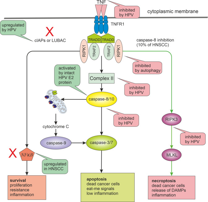
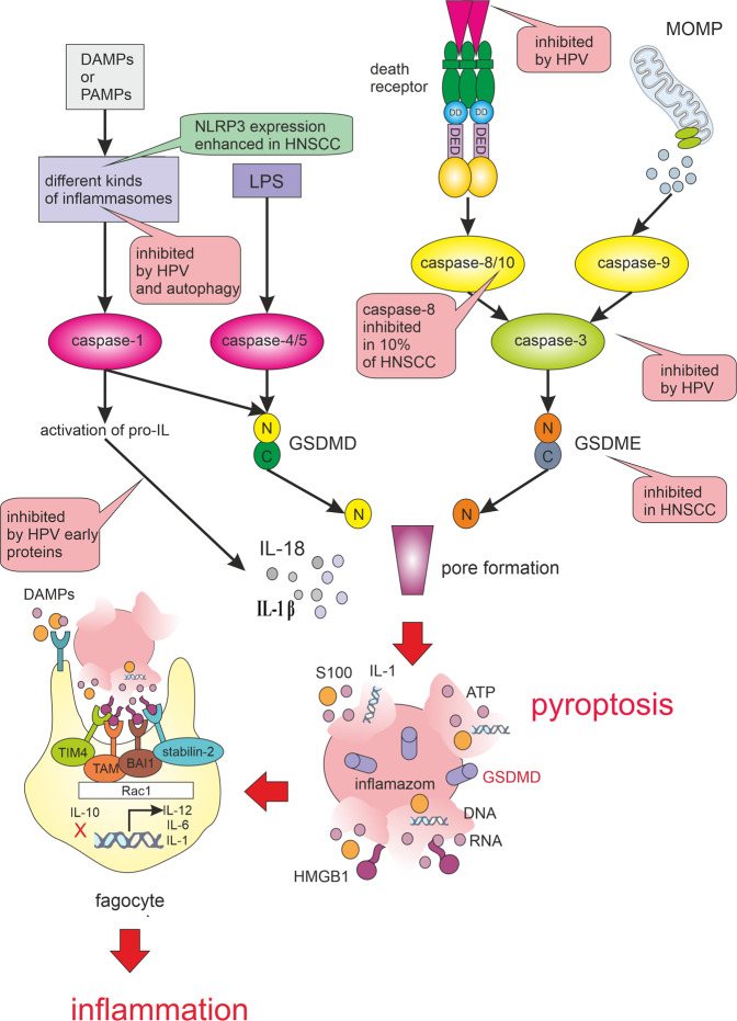
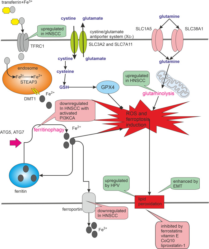
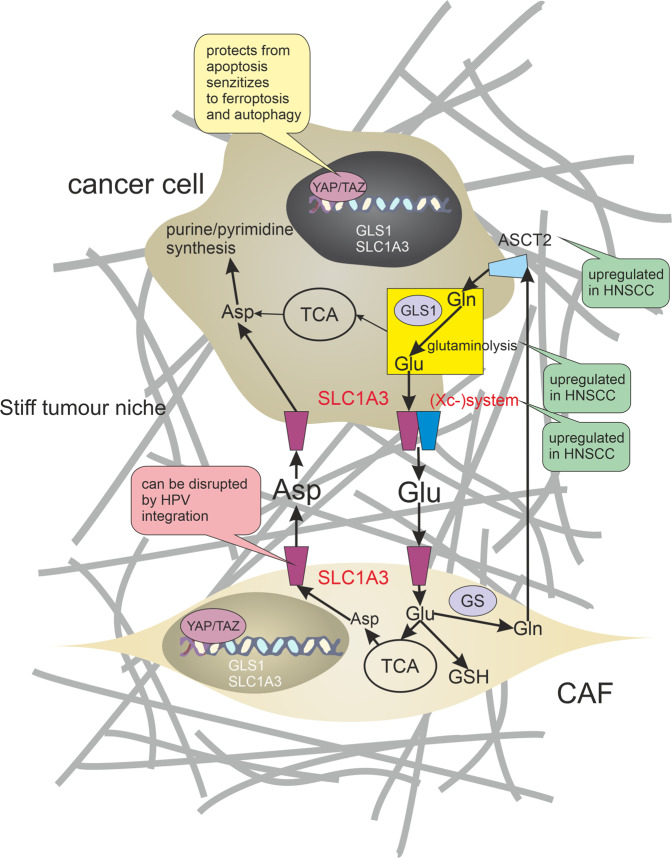
Many cancer therapies aim to trigger apoptosis in cancer cells. Nevertheless, the presence of oncogenic alterations in these cells and distorted composition of tumour microenvironment largely limit the clinical efficacy of this type of therapy. Luckily, scientific consensus describes about 10 different cell death subroutines with different regulatory pathways and cancer cells are probably not able to avoid all of cell death types at once. Therefore, a focused and individualised therapy is needed to address the specific advantages and disadvantages of individual tumours. Although much is known about apoptosis, therapeutic opportunities of other cell death pathways are often neglected. Molecular heterogeneity of head and neck squamous cell carcinomas (HNSCC) causing unpredictability of the clinical response represents a grave challenge for oncologists and seems to be a critical component of treatment response. The large proportion of this clinical heterogeneity probably lies in alterations of cell death pathways. How exactly cells die is very important because the predominant type of cell death can have multiple impacts on the therapeutic response as cell death itself acts as a second messenger. In this review, we discuss the different types of programmed cell death (PCD), their connection with HNSCC pathogenesis and possible therapeutic windows that result from specific sensitivity to some form of PCD in some clinically relevant subgroups of HNSCC.


Conflict of interest statement

The authors declare that they have no conflict of interest.

Figures

Fig. 1
Fig. 1. Alternative pathways of TNF signalling and their alterations in HNSCC.
TNF signalling can lead to different results. It depends on the post-translational modification and activation of key molecules such as caspase-8, RIPK1 and NF-κB. While active caspase-8 triggers apoptosis and suppresses necroptosis, its inactivity (e.g. by mutations) leads to necroptosis through RIPK3 activation. If the RIPK1 signal is modified by the addition of ubiquitin, cell death can be attenuated, and the cell receives a signal for survival and proliferation by the transcription factor NF-κB. NF-κB pathway inhibitors such as LUBAC inhibitors or SMAC mimetics can sensitise HNSCC cells to necroptosis or apoptosis. For successful anticancer treatment response, the pathway marked by red exes should be inhibited (inhibition of autophagy should be also beneficial for necroptosis triggering). The TNF signalling pathways are also significantly influenced by HPV infection and by the genetic background of HNSCC. Green bubbles indicate activation and pink inhibition of the process. LUBAC linear ubiquitin chain assembly complex, c-IAPs inhibitors of apoptosis, RIPK receptor-interacting serine/threonine–protein kinases, MLKL mixed lineage kinase domain-like pseudokinase.
Fig. 2
Fig. 2. Pyroptosis pathways. Pyroptosis is triggered when damage-associated molecular patterns (DAMPs) or pathogen-associated molecular patterns (PAMPs) activate the inflammasomes.
Active inflammasomes lead to the cleavage and activation of caspase-1. The activated caspase-1 cleaves Gasdermin D (GSDMD). The N-fragment of GSDMD form cell membrane pores resulting in pyroptosis. Activated caspase-1 also promotes the maturation and secretion of interleukins IL-1β and IL-18. When pathogen-derived lipopolysaccharide (LPS) binds to the precursor of caspase-4/5, it can also cause GSDMD cleavage and induction of pyroptosis. Another way to activate pyroptosis is caspase-3/Gasdermin E (GSDME) pathway. Caspase-3 can also be activated by mitochondrial outer membrane permeabilization (MOMP) and death receptor pathways. The activated caspase-3 then cleaves GSDME and produces GSDME N-fragments, The N-fragment of GSDME form cell membrane pores resulting in pyroptosis. Pyroptosis results in membrane rupture and the release of DAMPs that are detected by phagocyte receptors, such as TIM 4 (T cell immunoglobulin mucin receptor 4), BAI 1 (brain-specific angiogenesis inhibitor 1), stabilin-2 and TAM (Tryo3-Axl-Mer receptor). DAMPs recognition leads to the production of pro-inflammatory interleukins (e.g. IL-1, IL-6) and IL-12, which activates NK cells and induces the differentiation of naive CD4 T cells. The pyroptotic signalling pathways are significantly influenced by HPV infection and by the genetic background of HNSCC. Green bubbles indicate activation and pink inhibition of the process.
Fig. 3
Fig. 3. Ferroptosis. Ferroptosis is characterised by iron-induced lipid peroxidation.
The intracellular concentration of iron can be affected by the activity of transferrin, transferrin receptor and ferroportin, or by the release of iron from ferritin, which is often managed by ferritinophagy. HNSCC cells often manifest an increased intracellular iron concentration due to a high level of TFRC1 (transferrin receptor 1 responsible for cellular iron uptake) and a low abundance of ferroportin (responsible for iron efflux). The non-physiological degree of lipid peroxidation and the initiation of ferroptosis is prevented by glutathione and glutathione peroxidase 4 (GPX4) activity. Ferroptosis could be also prevented with chelating agents or vitamin E, which casts a bad light on the benefits of antioxidants in the treatment of some ferroptosis-prone cancers. Glutamine and glutaminolysis also play a crucial role in the activation of ferroptosis. PI3KCA is among the most frequently mutated and activated genes in HNSCC. PIK3CA activation can lead to increased mTOR activity and decreased autophagy. Consequently, the presence of PI3KCA activation may predispose these cancer cells to avoid autophagy, ferritinophagy and ferroptosis. The ferroptotic signalling pathways are significantly influenced by HPV infection and by the genetic background of HNSCC. Green bubbles indicate activation and pink inhibition of the process.
Fig. 4
Fig. 4. Metabolic symbiosis between cancer-associated fibroblasts and HNSCC cells.
HNSCC cells undergo numerous metabolic changes including increased glutaminolysis. Glutaminolysis produces large pools of intracellular glutamate. Upregulation of the cystine/glutamate antiporter ((Xc–) system) and excitatory amino acid transporter (SLC1A3) promotes aberrant glutamate (Glu) release from cancer cells. Increased stiffness of extracellular matrix during tumour progression induces cancer-associated fibroblasts (CAFs) to import glutamate (through SLC1A3) and release aspartate (Asp) and glutamine (Gln) supporting cancer cell purine/pyrimidine synthesis. Glutamine synthetase (GS) is often overexpressed in these CAFs. CAFs can also promote chemoresistance through the production of glutathione (GSH). Establishing of this metabolic symbiosis is coordinated by a YAP/TAZ-dependent mechanotransduction pathway. Green bubbles indicate activation and pink inhibition of the process.

References

    1. Lemaire F, et al. Differential expression profiling of head and neck squamous cell carcinoma (HNSCC) Br. J. Cancer. 2003;89:1940–1949. doi: 10.1038/sj.bjc.6601373. - DOI - PMC - PubMed
    1. Koch WM, Ridge JA, Forastiere A, Manola J. Comparison of clinical and pathological staging in head and neck squamous cell carcinoma: results from Intergroup Study ECOG 4393/RTOG 9614. Arch. Otolaryngol. Head. Neck Surg. 2009;135:851–858. doi: 10.1001/archoto.2009.123. - DOI - PMC - PubMed
    1. Keck MK, et al. Integrative analysis of head and neck cancer identifies two biologically distinct HPV and three non-HPV subtypes. Clin. Cancer Res. 2015;21:870–881. doi: 10.1158/1078-0432.CCR-14-2481. - DOI - PubMed
    1. Leemans CR, Snijders PJF, Brakenhoff RH. The molecular landscape of head and neck cancer. Nat. Rev. Cancer. 2018;18:269–282. doi: 10.1038/nrc.2018.11. - DOI - PubMed
    1. Wallace NA, Galloway DA. Manipulation of cellular DNA damage repair machinery facilitates propagation of human papillomaviruses. Semin. cancer Biol. 2014;26:30–42. doi: 10.1016/j.semcancer.2013.12.003. - DOI - PMC - PubMed

Publication types

MeSH terms

Substances