. 2021 Feb 18;11(2):39.
doi: 10.1038/s41408-021-00429-z.

Ibrutinib and venetoclax target distinct subpopulations of CLL cells: implication for residual disease eradication

Affiliations

Ibrutinib and venetoclax target distinct subpopulations of CLL cells: implication for residual disease eradication

Pin Lu et al. Blood Cancer J. .

Abstract

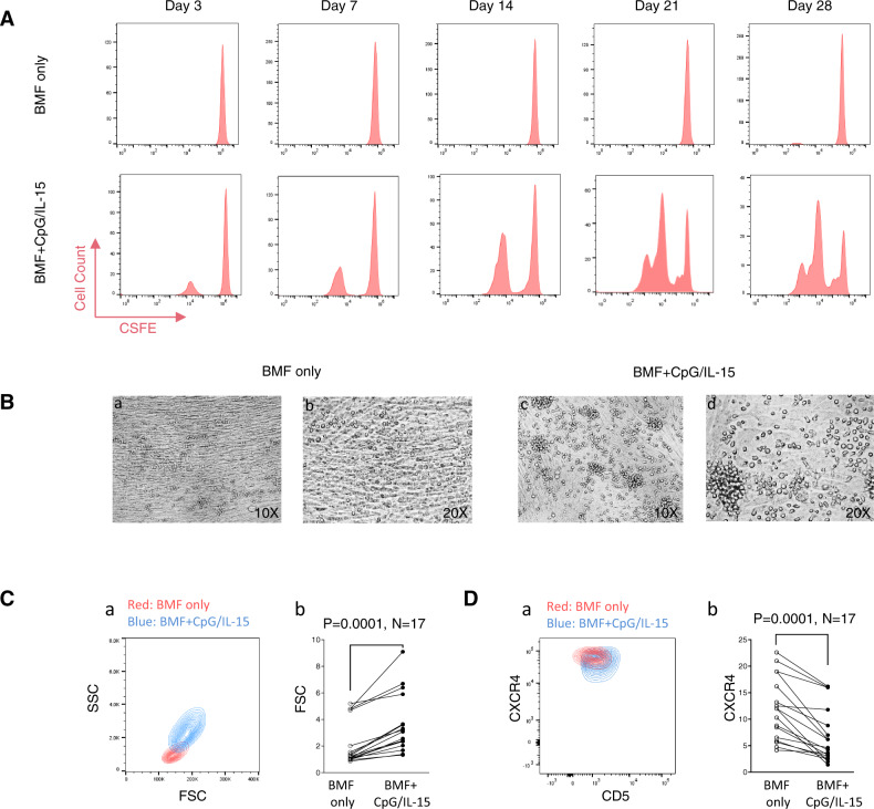
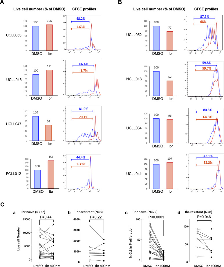
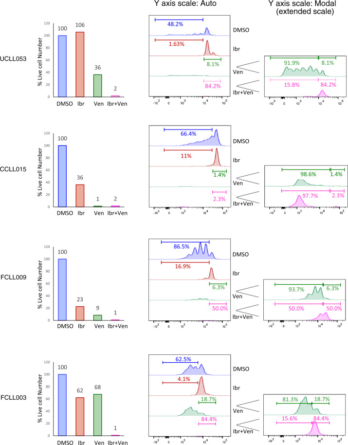
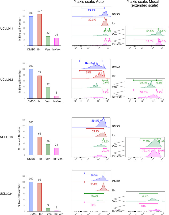
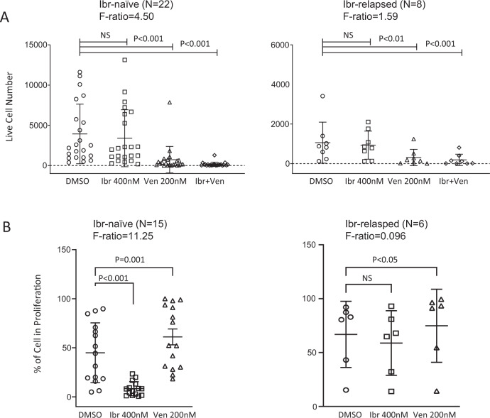
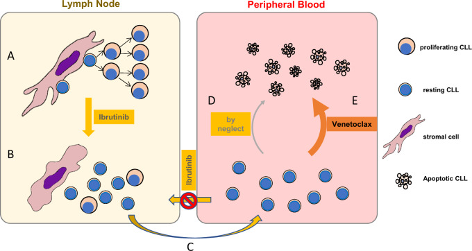
Ibrutinib inhibits Bruton tyrosine kinase while venetoclax is a specific inhibitor of the anti-apoptotic protein BCL2. Both drugs are highly effective as monotherapy against chronic lymphocytic leukemia (CLL), and clinical trials using the combination therapy have produced remarkable results in terms of rate of complete remission and frequency of undetectable minimal residual disease. However, the laboratory rationale behind the success of the drug combination is still lacking. A better understanding of how these two drugs synergize would eventually help develop other rational combination strategies. Using an ex vivo model that promotes CLL proliferation, we show that modeled ibrutinib proliferative responses, but not viability responses, correlate well with patients' actual clinical responses. Importantly, we demonstrate for the first time that ibrutinib and venetoclax act on distinct CLL subpopulations that have different proliferative capacities. While the dividing subpopulation of CLL responds to ibrutinib, the resting subpopulation preferentially responds to venetoclax. The combination of these targeted therapies effectively reduced both the resting and dividing subpopulations in most cases. Our laboratory findings help explain several clinical observations and contribute to the understanding of tumor dynamics. Additionally, our proliferation model may be used to identify novel drug combinations with the potential of eradicating residual disease.


Conflict of interest statement

Y.T., Speaker’s bureau for Novartis. S.M., Consultancy for AstraZeneca, Bioverativ, Genentech, Janssen, Kite and Pharmacyclics, Speaker’s bureau for AstraZeneca, Janssen and Pharmacyclics, and research funding from Abbvie, AstraZeneca, Beigene, Pharmacyclics, Juno and Novartis. N.K., advisory board for Pharmacyclics, Jessen and Seattle Genetics, and research funding from Bristol Meyer Squibb. Y.L.W., employment and stock ownership at Incyte Research Institute. All other authors declare no relevant conflicts of interest.

Figures

Fig. 1
Fig. 1. CLL cells actively divide for a long term in an ex vivo model.
Peripheral blood CLL cells from a patient were co-cultured with either BMF alone or with BMF+CpG/IL-15 stimulation. A Cell proliferation analyzed by flow cytometry. CFSE-labeled CLL cells were cultured with either BMF alone (upper) or with CpG (2 µg/mL) plus IL-15 (10 ng/mL) (lower). Duration of culture were indicated. B Light microscopy of CLL cells at Day 7 of the co-culture. CLL cells cultured with BMF alone are single small round cells laying on top of the BMF monolayer (a and b), while CLL cells with CpG/IL-15 stimulation appear larger and clustered (c and d). Microscopic magnifications are indicated. C Cell size was determined by forward scatter (FSC) on flow cytometry at Day 7 of the co-culture. SSC/FSC contour map between the two culture conditions is shown for one case (a) and for 17 cases (b). D Cell surface CXCR4 was measured by flow cytometry at Day 7 of the co-culture. CXCR4/CD5 contour map between the two culture conditions is shown for one case (a) and for 17 cases (b).
Fig. 2
Fig. 2. Only the dividing subpopulation of CLL responds to ibrutinib, which is largely consistent with patients’ actual clinical response.
CFSE-labeled CLL cells were cultured with BMF+CpG/IL-15 in the presence of DMSO or 400 nM of ibrutinib (Ibr). All analyses shown were conducted at day 7 of the co-culture by flow cytometry. A Live cell numbers (bar graphs) and CFSE profiles of 4 ibrutinib naïve/sensitive cases. Live cell numbers were normalize to the DMSO control (100%). Percentage of cells distributed in the dividing phases of the CFSE profiles is automatically calculated by the FlowJo software. B Live cell numbers and CFSE profiles of 4 ibrutinib resistant cases. C Aggregate results of cell viability and % of dividing CLL cells are shown for ibr naïve patients (a and c) as well as ibr resistant patients (b and d).
Fig. 3
Fig. 3. The resting subpopulation of CLL, as opposed to the dividing one, preferentially responds to venetoclax.
CFSE-labeled CLL cells were cultured with BMF+CpG/IL-15 in the presence of DMSO, 400 nM of ibr or 200 nM of venetoclax (ven). All analyses shown were conducted at day 7 of the co-culture by flow cytometry. A Live cell numbers (bar graphs) and CFSE profiles of two ibrutinib naïve/sensitive cases. Live cell numbers were normalize to the DMSO control (100%). Percentage of cells distributed in the dividing phases of the CFSE profiles is automatically calculated by the FlowJo software. Y axis scale-auto displays events on the same scale for all treatment conditions, and Y axis scale-Modal displays events on an extended scale to show low peaks under the condition of ventoclax treatment. B Live cell numbers and CFSE profiles of two ibrutinib resistant cases.
Fig. 4
Fig. 4. Combination of ibrutinib and venetoclax effectively eliminates both resting and dividing CLL subpopulations in ibrutinib-sensitive cases.
CFSE-labeled CLL cells were cultured with BMF+CpG/IL-15 in the presence of DMSO, 400 nM of ibr, 200 nM of venetoclax (ven) or both. All analyses shown were conducted at day 7 of the co-culture by flow cytometry. Live cell numbers (bar graphs) and CFSE profiles of four ibrutinib naïve/sensitive cases are shown. Live cell numbers were normalize to the DMSO control (100%). Percentage of cells distributed in the dividing phases of the CFSE profiles is automatically calculated by the FlowJo software. Y axis scale-auto displays events on the same scale for all treatment conditions, and Y axis scale-Modal displays events on an extended scale to show low peaks under the conditions of ventoclax or combination treatment.
Fig. 5
Fig. 5. Combination of ibrutinib and venetoclax effectively eliminates both resting and dividing CLL subpopulations in ibrutinib-resistant cases.
Live cell numbers and CFSE profiles of four ibrutinib-resistant cases are shown. Experimental conditions were identical to Fig. 4.
Fig. 6
Fig. 6. Effects of ibrutinib, venetoclax or combination on cell viability and cell proliferation.
A Aggregate results of cell viability for ibr naïve patients (left) and ibr resistant patients (right). B Aggregate results of percentage of the dividing CLL cells for ibr naïve patients (left) and ibrutinib-resistant patients (right). The combination was not depicted since analyses of the CFSE profiles became less meaningful in many cases due to the small number of residual live cells left after the combined treatment. p values and F ratios are indicated.
Fig. 7
Fig. 7. A schematic diagram of tumor cell dynamics under ibrutinib or venetoclax treatment.
A In lymph nodes, with close contact between tumor and stroma, CLL cells are well supported and proliferate. B With ibrutinibutinib treatment, adhesion to stromal cells is disrupted and proliferation is halted. C CLL cells are released to the periphery where they are also prevented from homing back to LN by the action of ibrutinibutinib on chemokine signaling. D In the periphery, without the supporting tumor microenvironment, the cells become non-proliferating/resting and die by neglect over a long period of time. E In the presence of venetoclax, however, these resting cells are subjective to active killing, causing tumor lysis syndrome and disease improvement.

References

    1. Rozman C, Montserrat E. Chronic lymphocytic leukemia. N. Engl. J. Med. 1995;333:1052–1057. doi: 10.1056/NEJM199510193331606. - DOI - PubMed
    1. Chiorazzi N, Rai KR, Ferrarini M. Chronic lymphocytic leukemia. N. Engl. J. Med. 2005;352:804–815. doi: 10.1056/NEJMra041720. - DOI - PubMed
    1. Byrd JC, et al. Targeting BTK with ibrutinib in relapsed chronic lymphocytic leukemia. N. Engl. J. Med. 2013;369:32–42. doi: 10.1056/NEJMoa1215637. - DOI - PMC - PubMed
    1. Roberts AW, et al. Targeting BCL2 with venetoclax in relapsed chronic lymphocytic leukemia. N. Engl. J. Med. 2016;374:311–322. doi: 10.1056/NEJMoa1513257. - DOI - PMC - PubMed
    1. Roberts AW, et al. Efficacy of venetoclax in relapsed chronic lymphocytic leukemia is influenced by disease and response variables. Blood. 2019;134:111–122. doi: 10.1182/blood.2018882555. - DOI - PMC - PubMed

Publication types

MeSH terms

LinkOut - more resources