Review
. 2021 Feb 18;7(1):13.
doi: 10.1038/s41572-021-00248-3.

Duchenne muscular dystrophy

Affiliations
Review

Duchenne muscular dystrophy

Dongsheng Duan et al. Nat Rev Dis Primers. .

Abstract

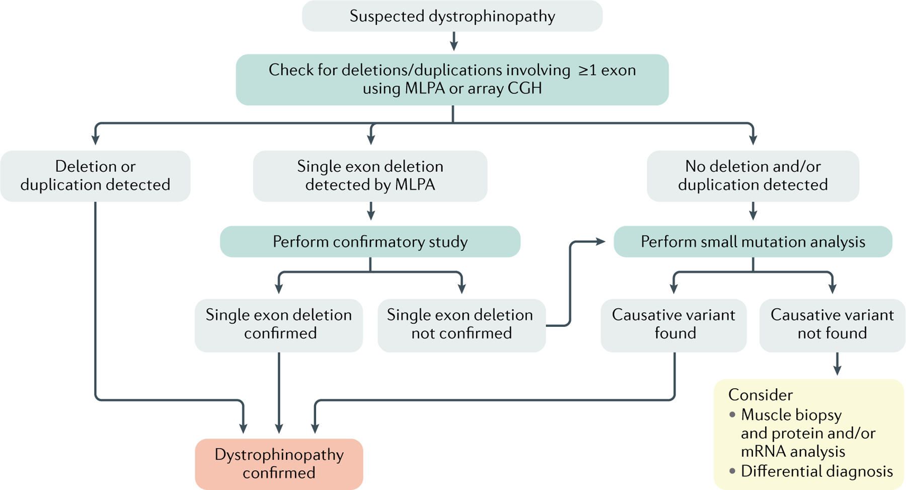
Duchenne muscular dystrophy is a severe, progressive, muscle-wasting disease that leads to difficulties with movement and, eventually, to the need for assisted ventilation and premature death. The disease is caused by mutations in DMD (encoding dystrophin) that abolish the production of dystrophin in muscle. Muscles without dystrophin are more sensitive to damage, resulting in progressive loss of muscle tissue and function, in addition to cardiomyopathy. Recent studies have greatly deepened our understanding of the primary and secondary pathogenetic mechanisms. Guidelines for the multidisciplinary care for Duchenne muscular dystrophy that address obtaining a genetic diagnosis and managing the various aspects of the disease have been established. In addition, a number of therapies that aim to restore the missing dystrophin protein or address secondary pathology have received regulatory approval and many others are in clinical development.


Conflict of interest statement

Competing interests

D.D. is a member of the scientific advisory board for Solid Biosciences and an equity holder of Solid Biosciences. D.D. is an inventor on patents licensed to various companies. D.D. has served as an ad hoc consultant for 4DMT, Decibel Therapeutics, Evox, Primary Insight, Vida Ventures, Global Guidepoint and GLG consultancy in the past 3 years. The lab of D.D. has received research support from Solid Biosciences and Edgewise Therapeutics in the past 3 years. N.G. has received compensation as member of scientific boards or as speaker at symposia from Sarepta, Pfizer, Italpharmaco and PTC Therapeutics. S.T. has patents on sequences for exon skip by antisense nucleic acids as a member of NCNP together with Nippon Shinyaku. As principal inventor of these patents, S.T. is entitled to a share of royalties. S.T. discloses being an ad hoc consultant for Ono Pharmaceutical, Daiichisankyo, Asahikasei Pharma, Teijin Pharma, AGADA Biosciences, and Wave and being a member of the scientific advisory boards of Nippon Shinyaku, Taiho Pharma and Sarepta therapeutics. S.T. received speaker honoraria from Japan Health Science Foundation and Astellas Pharma and has also received research supports from Taiho Pharma, Daiichisankyo, Nippon Shinyaku, Takeda Pharmaceutical and the Noguchi Institute in the past 3 years. E.M. is a Principal Investigator in clinical trials and advisory board member for Sarepta, Santhera, PTC, Roche, Italfarmaco, NS Pharma and Pfizer. A.A.-R. discloses being employed by Leiden University Medical Center (LUMC), which has patents on exon-skipping technology, some of which has been licensed to BioMarin and subsequently sublicensed to Sarepta. As co-inventor of some of these patents, A.A.-R. is entitled to a share of royalties. A.A.-R. further discloses being an ad hoc consultant for PTC Therapeutics, Sarepta Therapeutics, CRISPR Therapeutics, Summit PLC, Alpha Anomeric, BioMarin Pharmaceuticals Inc., Eisai, Astra Zeneca, Santhera, Audentes, Global Guidepoint and GLG consultancy, Grunenthal, Wave, and BioClinica, having been a member of the Duchenne Network Steering Committee (BioMarin), and being a member of the scientific advisory boards of ProQR, Hybridize Therapeutics, Silence Therapeutics, Sarepta Therapeutics and Philae Pharmaceuticals. Remuneration for these activities is paid to LUMC. LUMC also received speaker honoraria from PTC Therapeutics and BioMarin Pharmaceuticals and funding for contract research from Italpharmaco and Alpha Anomeric. Project funding is received from Sarepta Therapeutics.

Figures

Fig. 1 |
Fig. 1 |. Schematic depiction of DMD and dystrophin protein.
a | The ~2.4 Mb full-length DMD gene contains eight promoters and 79 exons. Three upstream promoters (Dp427b, Dp427m and Dp427p) produce the ~11.4 kb full-length cDNA and the 427 kDa full-length dystrophin protein. Four internal promoters (Dp260, Dp140, Dp116 and Dp71) generate N-terminal truncated non-muscle isoforms of dystrophin. Alternative splicing at the 3’-end and alternative polyadenylation (the addition of a poly(A) tail to RNA) yield additional isoforms of dystrophin such as Dp40. The full-length protein generated from Dp427m is the primary muscle isoform. b | Full-length dystrophin can be divided into four major domains, including the N-terminal F-actin-binding domain (ABD; encoded by exons 1–8), rod (R; encoded by exons 8–64), cysteine-rich (CR; encoded by exons 64–70) and C-terminal (CT; encoded by exons 71–79) domains. The rod domain can be further divided into 24 spectrin-like repeats and four interspersed hinges. c | In patients with Duchenne muscular dystrophy (DMD), protein production is prematurely truncated and the resulting protein is not functional. This leads to the loss of connections between the cytoskeleton and the extracellular matrix. d | By contrast, in patients with Becker muscular dystrophy, a partially functional dystrophin is produced that contains the crucial domains required to connect to F-actin and the extracellular matrix.
Fig. 2 |
Fig. 2 |. Healthy muscle and DMD muscle histology.
Cross-sectional staining of healthy muscle (panels ad) and skeletal muscle from a patient with Duchenne muscular dystrophy (DMD; panels eh). Haematoxylin and eosin (HE) staining shows centrally nucleated myofibers, inflammatory cell infiltration, variable myofiber size, and endomysium and perimysium connective tissue deposition (panels a and e). Masson trichrome (MT) staining shows increased fibrosis (blue staining) in a patient with DMD compared with healthy muscle (panels b and f). Immunofluorescence labelling of dystrophin and laminin shows a lack of dystrophin in a patient with DMD compared with healthy muscle (panels c and g) and variation in myofiber size in DMD muscle (panels d and h).
Fig. 3 |
Fig. 3 |. Dystrophin and binding partners.
Dystrophin binding partners can be categorized as cytoskeletal proteins, transmembrane proteins, extracellular proteins, cytosolic signalling and scaffolding proteins, endocytic proteins, and cardiac-specific interacting proteins. ABD, actin-binding domain; CNS, central nervous system; CR, cysteine-rich domain; CT, C-terminal domain; DG, dystroglycan; NCX, sodium-calcium exchanger; nNOS, neuronal nitric oxide synthase; NOX2, NADPH oxidase 2; PMCA, plasma membrane calcium ATPase.
Fig. 4 |
Fig. 4 |. Diagnostic decision tree to confirm the genetic diagnosis of dystrophinopathies.
Boys with typical symptoms of a dystrophinopathy and increased plasma creatine kinase (CK) levels should undergo genetic testing to confirm the diagnosis. Initially, multiplex ligation-dependent probe amplification (MLPA) or array comparative genome hybridization (CGH) should be used to identify causative DMD variants; however, if a causative variant is not identified (which occurs in ~25% of patients), small mutation analysis should be used. Boys without an identified causative variant after small mutation analysis should be referred for muscle biopsy and protein or mRNA analysis.
Fig. 5 |
Fig. 5 |. Dystrophin-restoring approaches.
a | Micro-dystrophin gene therapy. To accommodate the limited capacity of adeno-associated viral vectors, cDNA constructs encoding only the most crucial parts of dystrophin have been generated to enable the expression of micro-dystrophins. b | Exon skipping. Antisense oligonucleotides (AONs) are used to target a specific exon during pre-mRNA splicing. The AON hides the targeted exon from the splicing machinery causing it to be skipped, thus restoring the reading frame, allowing the production of a Becker-like dystrophin. c | Genome editing. Guide RNAs are used to direct Cas9 to target regions in the DNA to delete an exon from the gene, thereby restoring the reading frame of mRNA produced from this gene, allowing the production of Becker-like dystrophin. d | Stop codon readthrough. For patients with nonsense mutations (indicated by an asterisk), compounds can suppress the premature stop codon use, facilitating instead the inclusion of an amino acid. Thus, a full-length dystrophin can be produced. ABD, actin-binding domain; CR, cysteine-rich domain; CT, C-terminal domain.

References

    1. Mercuri E, Bonnemann CG & Muntoni F Muscular dystrophies. Lancet 394, 2025–2038 (2019).

      Comprehensive overview of the clinical and genetic aspects of muscular dystrophies.

    1. Aartsma-Rus A, Van Deutekom JCT, Fokkema IF, Van Ommen GJB & Den Dunnen JT Entries in the Leiden Duchenne muscular dystrophy mutation database: an overview of mutation types and paradoxical cases that confirm the reading-frame rule. Muscle Nerve 34, 135–144 (2006). - PubMed
    1. Monaco AP, Bertelson CJ, Liechti-Gallati S, Moser H & Kunkel LM An explanation for the phenotypic differences between patients bearing partial deletions of the DMD locus. Genomics 2, 90–95 (1988). - PubMed
    1. García-Rodríguez R et al. Premature termination codons in the DMD gene cause reduced local mRNA synthesis. Proc. Natl Acad. Sci. USA 117, 16456–16464 (2020). - PMC - PubMed
    1. Mendell JR et al. Evidence-based path to newborn screening for Duchenne muscular dystrophy. Ann. Neurol 71, 304–313 (2012). - PubMed

Publication types

MeSH terms