. 2021 Feb 18;11(1):4215.
doi: 10.1038/s41598-021-83762-4.

Sophisticated viral quasispecies with a genotype-related pattern of mutations in the hepatitis B X gene of HBeAg-ve chronically infected patients

Affiliations

Sophisticated viral quasispecies with a genotype-related pattern of mutations in the hepatitis B X gene of HBeAg-ve chronically infected patients

Maria Francesca Cortese et al. Sci Rep. .

Abstract

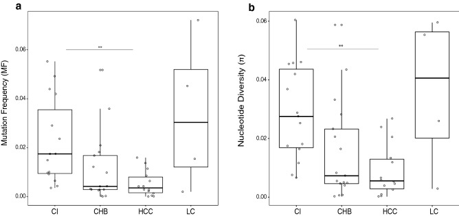
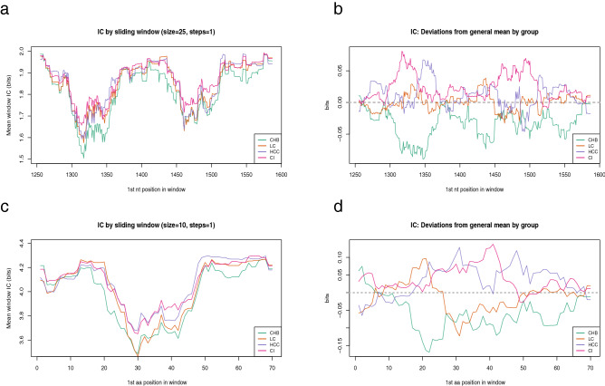
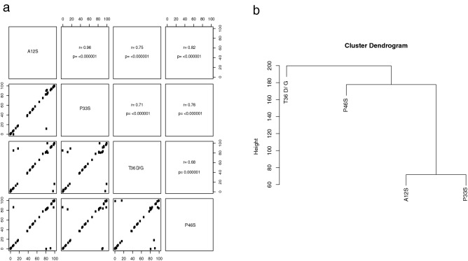
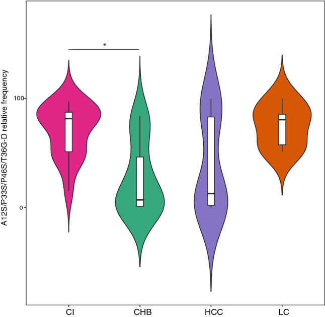
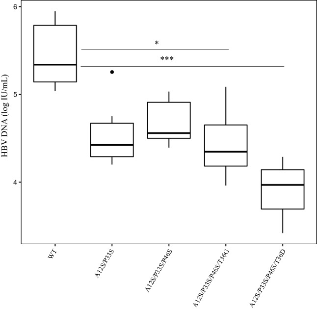
Patients with HBeAg-negative chronic infection (CI) have not been extensively studied because of low viremia. The HBx protein, encoded by HBX, has a key role in viral replication. Here, we analyzed the viral quasispecies at the 5' end of HBX in CI patients and compared it with that of patients in other clinical stages. Fifty-eight HBeAg-negative patients were included: 16 CI, 19 chronic hepatitis B, 16 hepatocellular carcinoma and 6 liver cirrhosis. Quasispecies complexity and conservation were determined in the region between nucleotides 1255 and 1611. Amino acid changes detected were tested in vitro. CI patients showed higher complexity in terms of mutation frequency and nucleotide diversity and higher quasispecies conservation (p < 0.05). A genotype D-specific pattern of mutations (A12S/P33S/P46S/T36D-G) was identified in CI (median frequency, 81.7%), which determined a reduction in HBV DNA release of up to 1.5 log in vitro. CI patients showed a more complex and conserved viral quasispecies than the other groups. The genotype-specific pattern of mutations could partially explain the low viremia observed in these patients.


Conflict of interest statement

Josep Gregori is an employee of Roche Diagnostics, SL. No competing interests are reported for the other authors.

Figures

Figure 1
Figure 1
Mutation frequency (Mf) and population nucleotide diversity (π) comparisons. Boxplots show comparisons of Mf (a) and π (b) values between the 4 clinical groups. Each dot represents a single patient. Asterisks above the boxes denote specific p values (2 asterisks < 0.01) obtained by performing the Kruskal–Wallis test plus Dunn posthoc test and adjusted with the Bonferroni correction. CHB chronic hepatitis B; CI chronic infection, HCC hepatocellular carcinoma, LC liver cirrhosis.
Figure 2
Figure 2
Intergroup variability of the information content. (a) The sliding window analysis is the result of the mean information content (bits) of the 25-nt or 10-aa long windows with a displacement between them of 1-nt or 1-aa obtained by multiple alignments of all the QS haplotypes. A. Sliding window analysis of nt sequences in the various clinical groups. Each line displays a specific clinical group (CHB in green, LC in orange, HCC in purple, and CI in pink). (b) Representation of the intergroup variability in nt conservation. Each line indicates a group of patients. The information content general mean is highlighted with a grey dotted line (positive deviation indicates greater conservation, whereas negative deviation indicates lesser conservation). (c) Sliding window analysis of aa sequences in the various clinical groups. (d) Intergroup variability in aa-conservation. Grey dotted line indicates the general mean of information content.
Figure 3
Figure 3
Pattern of mutations in genotype D haplotypes. (a) Spearman correlation between the frequencies of the amino acid changes highlighted in genotype D haplotypes. Each dot represents the change’s frequency in each patient. Rho and p values are reported. (b) Hierarchical cluster dendrogram of the observed mutations. The y-axis is a measure of closeness of an individual mutation or a cluster.
Figure 4
Figure 4
Frequency of the genotype D-specific mutational pattern. The violin plot compares the frequency of the mutational pattern (A12S/P33S/P46S/T36G-D) between the clinical groups. The internal white bar represents the interquartile range with the median in a black horizontal line, whereas the vertical line shows the 95% confidential interval. The violin shape indicates the probability density, in which wider sections indicate a higher probability that a patient will have a given frequency of the mutational pattern. The p value was determined by performing the Kruskal–Wallis test plus and the posthoc Dunn test, and is reported as asterisks (1 asterisk, < 0.05).
Figure 5
Figure 5
In vitro HBV expression in the presence of HBx mutations. The boxplot represents HBV particle release (HBV DNA in log IU/mL) in cell supernatants at 5dpt in the presence of wild type (wt) or mutated HBV monomers. The Kruskal–Wallis plus Dunn test was implemented and the p values obtained are reported as asterisks, where the number of asterisks identifies a specific p value interval (1 asterisk, < 0.05 and 3, < 0.0001). Results are the median of three experiments performed in duplicate.

References

    1. Okamoto H, et al. Typing hepatitis B virus by homology in nucleotide sequence: Comparison of surface antigen subtypes. J. Gen. Virol. 1988;69:2575–2583. doi: 10.1099/0022-1317-69-10-2575. - DOI - PubMed
    1. Lin Y-Y, et al. New insights into the evolutionary rate of hepatitis B virus at different biological scales. J. Virol. 2015;89:3512–3522. doi: 10.1128/JVI.03131-14. - DOI - PMC - PubMed
    1. Belloni L, et al. Nuclear HBx binds the HBV minichromosome and modifies the epigenetic regulation of cccDNA function. Proc. Natl. Acad. Sci. USA. 2009;106:19975–19979. doi: 10.1073/pnas.0908365106. - DOI - PMC - PubMed
    1. Lucifora J, et al. Hepatitis B virus X protein is essential to initiate and maintain virus replication after infection. J. Hepatol. 2011;55:996–1003. doi: 10.1016/j.jhep.2011.02.015. - DOI - PubMed
    1. Pollicino T, et al. Hepatitis B virus replication is regulated by the acetylation status of hepatitis B virus cccDNA-bound H3 and H4 histones. Gastroenterology. 2006;130:823–837. doi: 10.1053/j.gastro.2006.01.001. - DOI - PubMed

Publication types

LinkOut - more resources