. 2021 Feb 10:13:1257-1270.
doi: 10.2147/CMAR.S290645. eCollection 2021.

Circ_0056285 Regulates Proliferation, Apoptosis and Glycolysis of Osteosarcoma Cells via miR-1244/TRIM44 Axis

Affiliations

Circ_0056285 Regulates Proliferation, Apoptosis and Glycolysis of Osteosarcoma Cells via miR-1244/TRIM44 Axis

Shousong Huo et al. Cancer Manag Res. .

Abstract

Background: Osteosarcoma (OS) is a common malignant bone cancer that occurs in adolescents and children. Circular RNAs (circRNAs) are important regulators of tumorigenesis and development. This study aimed to explore the role and molecular basis of circ_0056285 in OS.

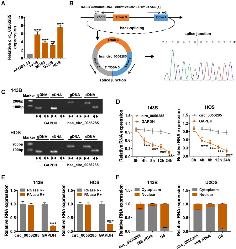
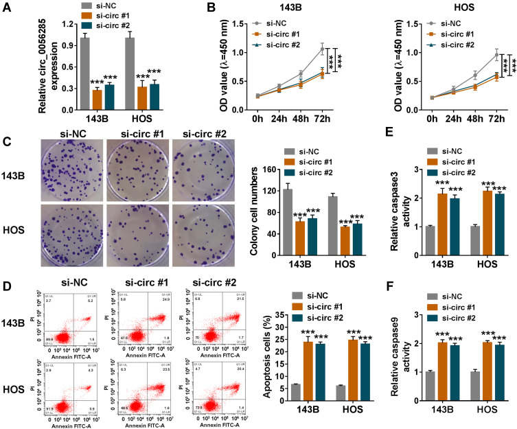
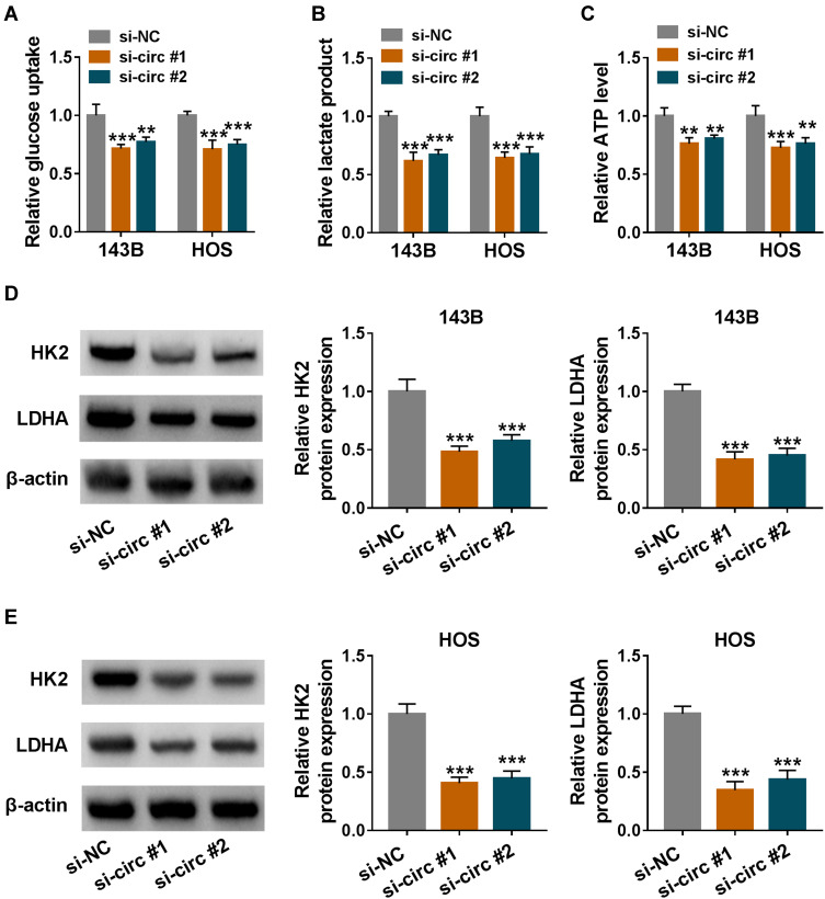
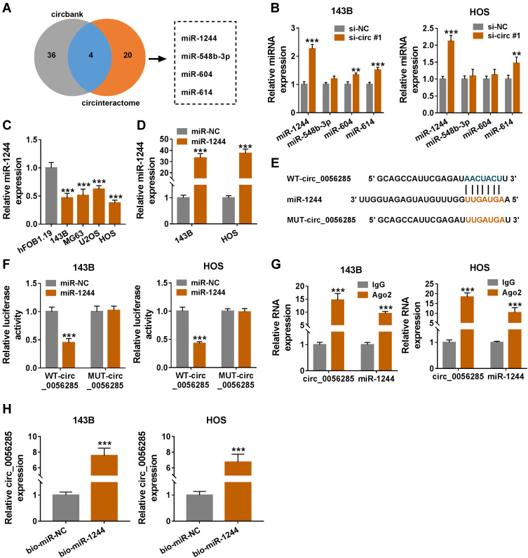
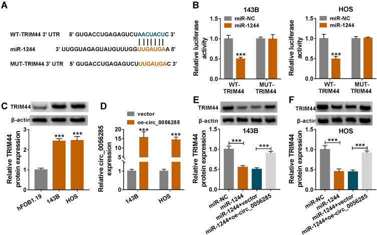
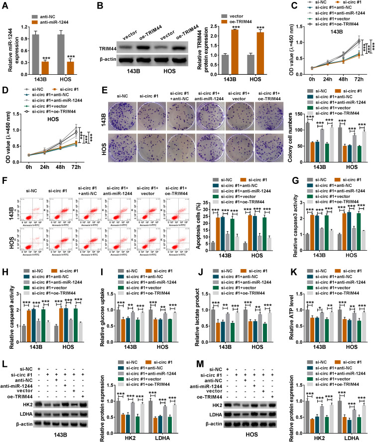
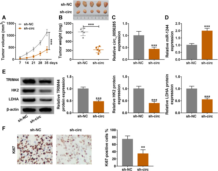
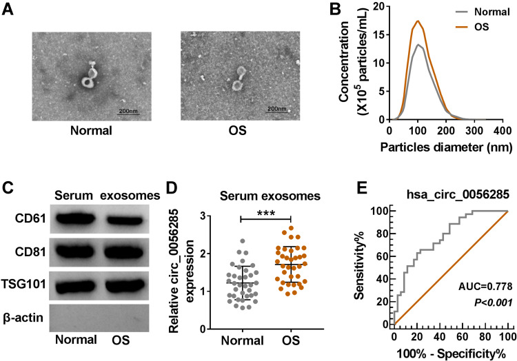
Methods: The levels of circ_0056285, miR-1244 and tripartite motif containing 44 (TRIM44) were determined by quantitative real-time polymerase chain reaction or Western blot assay. Cell proliferation was evaluated by Cell Counting Kit-8 (CCK-8) assay and colony formation assay. Cell apoptosis was assessed by flow cytometry and caspase 3and caspase 9 activity assay kits. Glucose uptake, lactate product and ATP level were examined using commercial kits. Hexokinase II (HK2) and lactate dehydrogenase A (LDHA) levels were measured by Western blot assay. The interaction among circ_0056285, miR-1244 and TRIM44 was confirmed by dual-luciferase reporter assay, RNA immunoprecipitation assay or RNA pull-down assay. Xenograft experiment was conducted to explore tumor growth in vivo. Exosomes were identified by transmission electron microscope (TEM), nanoparticle tracking analysis (NTA) and Western blot. The diagnostic value of exosomal circ_0056285 was evaluated by receiver operating characteristic (ROC) curve.

Results: Circ_0056285 and TRIM44 were up-regulated, and miR-1244 was down-regulated in OS tissues and cells. Circ_0056285 silencing inhibited proliferation and glycolysis and promoted apoptosis in OS cells. Also, circ_0056285 knockdown hindered proliferation and accelerated apoptosis in OS cells by regulating miR-1244/TRIM44 axis. Circ_0056285 depletion impeded tumor growth in vivo. Furthermore, ROC curve showed that circ_0056285 might be a diagnostic biomarker in OS.

Conclusion: Circ_0056285 facilitated OS progression by sponging miR-1244 and increasing TRIM44 expression, providing a promising therapeutic target for OS.

Keywords: TRIM44; circ_0056285; miR-1244; osteosarcoma; progression.


Conflict of interest statement

The authors declare that they have no conflict of interest.

Figures

Figure 1
Figure 1
Expression and validation of circ_0056285 in OS cells. (A) The expression of circ_0056285 in hFOB1.19 and OS cell lines (143B, MG63, U2OS and HOS) was measured by qRT-PCR. (B) The schematic illustration showed the origin of circ_0056285 and the result of Sanger sequencing. (C) Circ_0056285 was amplified by divergent primers in cDNA but not gDNA. (D) 143B and HOS cells were treated with Actinomycin D, and the expression of circ_0056285 and GAPDH was examined by qRT-PCR at different times. (E) The levels of circ_0056285 and GAPDH were detected in 143B and HOS cells treated with or without RNase R. (F) The levels of 18S rRNA, U6 and circ_0056285 in nuclear and cytoplasmic fractions were evaluated by qRT-PCR. **P < 0.01, ***P < 0.001.
Figure 2
Figure 2
Circ_0056285 knockdown inhibited proliferation and promoted apoptosis in OS cells. 143B and HOS cells were introduced with si-NC, si-circ#1 or si-circ#2. (A) The knockdown efficiency was assessed by qRT-PCR. (B and C) Cell proliferative capacity was evaluated by CCK-8 assay and colony formation assay. (D) Cell apoptosis was detected by flow cytometry. (E and F) The activities of caspase 3 and caspase 9 were examined using commercial kits. ***P < 0.001.
Figure 3
Figure 3
Circ_0056285 silencing suppressed the glycolysis of OS cells. 143B and HOS cells were transfected with si-NC, si-circ#1 or si-circ#2. (A and B) After transfection for 48 h, glucose uptake and lactate product were detected. (C) ATP level was measured using ATP colorimetric assay kit. (D and E) The levels of glycolysis-related proteins (HK2 and LDHA) were examined by Western blot assay. **P < 0.01, ***P < 0.001.
Figure 4
Figure 4
Circ_0056285 directly interacted with miR-1244. (A) Venn diagram showed miRNAs bound to circ_0056285 in Circbank and circinteractome databases. (B) The levels of four miRNAs (miR-1244, miR-548b-3p, miR-604 and miR-614) were tested in 143B and HOS cells transfected with si-NC or si-circ#1. (C) MiR-1244 expression in hFOB1.19 and OS cell lines (143B, MG63, U2OS and HOS) was evaluated using qRT-PCR. (D) The expression of miR-1244 was tested in 143B and HOS cells transfected with miR-1244 or miR-NC. (E) The predicted binding sites of circ_0056285 and miR-1244 were exhibited. (F) 143B and HOS cells were co-transfected with WT-circ_0056285 or MUT-circ_0056285 and miR-1244 or miR-NC, and luciferase activity was detected by dual-luciferase reporter assay. (G and H) The interaction between circ_0056285 and miR-1244 was further verified using RIP assay and RNA pull-down assay. **P < 0.01, ***P < 0.001.
Figure 5
Figure 5
Circ_0056285 sponged miR-1244 to regulate TRIM44 expression. (A) The putative binding sites for miR-1244 and TRIM44 3ʹUTR were shown. (B) The relationship between miR-1244 and TRIM44 was validated by dual-luciferase reporter assay. (C) TRIM44 protein level was detected in hFOB1.19 and OS cell lines (143B and HOS) using Western blot assay. (D) Circ_0056285 expression was measured in 143B and HOS cells transfected with vector or oe-circ_0056285. (E and F) 143B and HOS cells were introduced with miR-NC, miR-1244, miR-1244+vector or miR-1244+oe-circ_0056285, and TRIM44 protein expression was examined by Western blot assay. ***P < 0.001.
Figure 6
Figure 6
Inhibition of miR-1244 or overexpression of TRIM44 reversed the effect of circ_0056285 depletion on OS progression. (A) The expression of miR-1244 was detected in 143B and HOS cells transfected with anti-NC or anti-miR-1244. (B) TRIM44 protein level was examined in 143B and HOS cells introduced with vector or oe-TRIM44. (CM) 143B and HOS cells were transfected with si-NC, si-circ#1, si-circ#1+anti-NC, si-circ#1+anti-miR-1244, si-circ#1+vector or si-circ#1+oe-TRIM44, respectively. (C and D) Cell viability was assessed by CCK-8 assay. (E) The number of colonies was detected by colony formation assay. (F) Cell apoptosis was monitored by flow cytometry. (G and H) Caspase 3 and caspase 9 activities were tested by commercial kits. (IK) After 48 h of transfection, glucose uptake, lactate product and ATP level were detected. (L and M) Western blot assay was utilized to detect the protein levels of HK2 and LDHA. *P < 0.05, **P < 0.01, ***P < 0.001.
Figure 7
Figure 7
Circ_0056285 knockdown blocked tumor growth in vivo. 143B cells stably transfected with sh-NC or sh-circ were subcutaneously injected into the right-back of nude mice. (A) After 7 days, the growth curve of xenograft tumor was plotted. (B) After 35 days, the mice were sacrificed, and the tumors were weighed. (C and D) The levels of circ_0056285 and miR-1244 were measured using qRT-PCR. (E) The protein levels of TRIM44, HK2 and LDHA were detected by Western blot. (F) IHC assay was used to analyze Ki67 in xenograft tumor tissues. **P < 0.01, ***P < 0.001.
Figure 8
Figure 8
Circ_0056285 was highly expressed in serum exosomes of OS patients. (A) Representative images of serum exosomes were examined by TEM. (B) Size distribution was analyzed by NTA. (C) Exosome markers (CD61, CD81 and TSG101) were detected by Western blot. (D) Circ_0056285 expression in serum exosomes of OS patients (n=35) and healthy volunteers (n=35) was measured by qRT-PCR. (E) ROC curve was used to evaluate the diagnostic value of exosomal hsa_circ_0056285 in OS. ***P < 0.001.

References

    1. Whelan JS, Davis LE. Osteosarcoma, chondrosarcoma, and chordoma. J Clin Oncol. 2018;36(2):188–193. doi:10.1200/JCO.2017.75.1743 - DOI - PubMed
    1. Luetke A, Meyers PA, Lewis I, Juergens H. Osteosarcoma treatment–where do we stand? A state of the art review. Cancer Treat Rev. 2014;40(4):523–532. doi:10.1016/j.ctrv.2013.11.006 - DOI - PubMed
    1. Chen LL. The biogenesis and emerging roles of circular RNAs. Nat Rev Mol Cell Biol. 2016;17(4):205–211. doi:10.1038/nrm.2015.32 - DOI - PubMed
    1. Shang Q, Yang Z, Jia R, Ge S. The novel roles of circRNAs in human cancer. Mol Cancer. 2019;18(1):6. doi:10.1186/s12943-018-0934-6 - DOI - PMC - PubMed
    1. Wang C, Ren M, Zhao X, Wang A, Wang J. Emerging roles of circular RNAs in osteosarcoma. Med Sci Monit. 2018;24:7043–7050. doi:10.12659/MSM.912092 - DOI - PMC - PubMed

LinkOut - more resources