. 2021 Feb 2:11:623324.
doi: 10.3389/fimmu.2020.623324. eCollection 2020.

T-Bet Controls Cellularity of Intestinal Group 3 Innate Lymphoid Cells

Affiliations

T-Bet Controls Cellularity of Intestinal Group 3 Innate Lymphoid Cells

Jan-Hendrik Schroeder et al. Front Immunol. .

Abstract

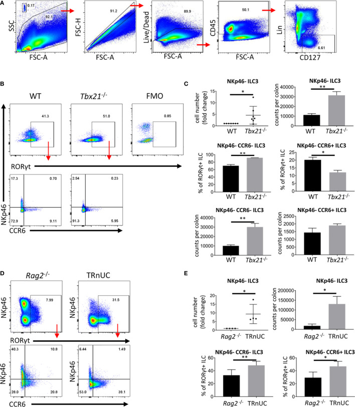
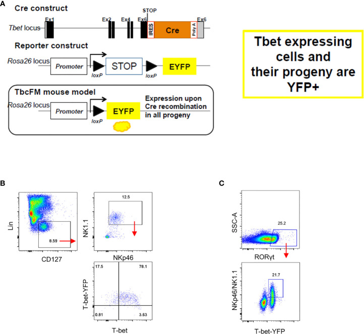
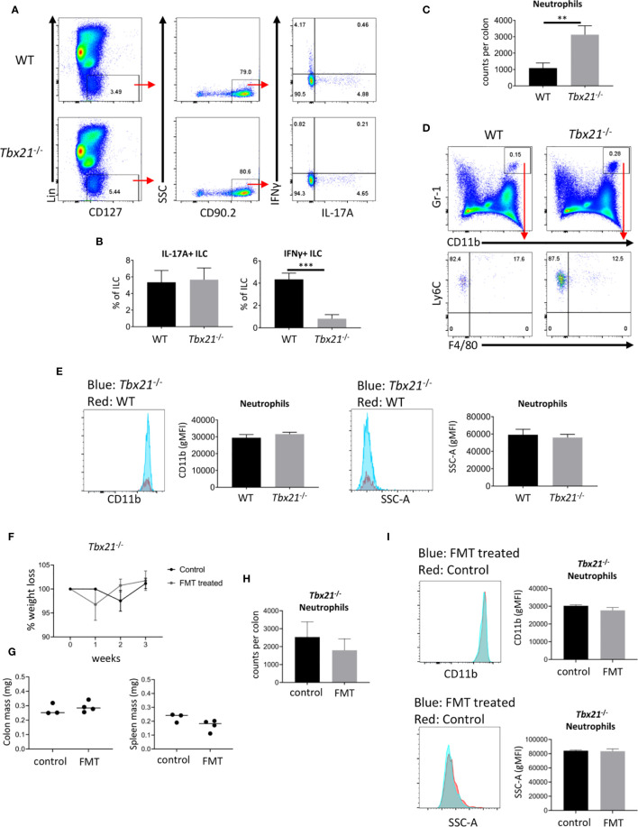
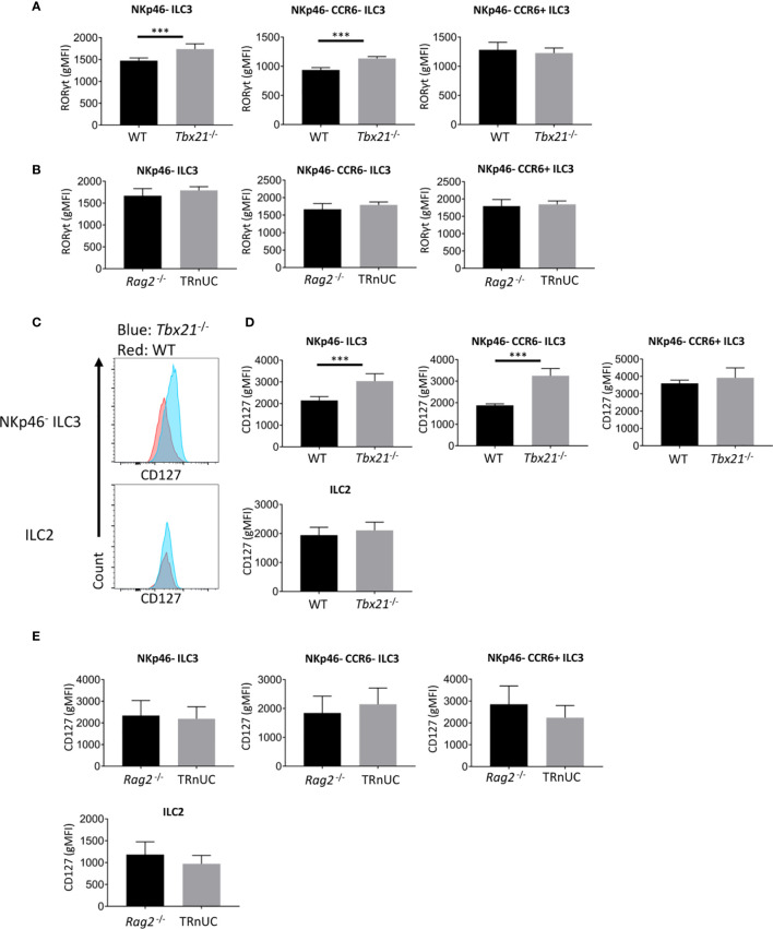
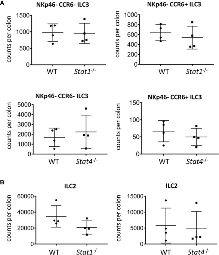
Innate lymphoid cells (ILC) play a significant immunological role at mucosal surfaces such as the intestine. T-bet-expressing group 1 innate lymphoid cells (ILC1) are believed to play a substantial role in inflammatory bowel disease (IBD). However, a role of T-bet-negative ILC3 in driving colitis has also been suggested in mouse models questioning T-bet as a critical factor for IBD. We report here that T-bet deficient mice had a greater cellularity of NKp46-negative ILC3 correlating with enhanced expression of RORγt and IL-7R, but independent of signaling through STAT1 or STAT4. We observed enhanced neutrophilia in the colonic lamina propria (cLP) of these animals, however, we did not detect a greater risk of T-bet-deficient mice to develop spontaneous colitis. Furthermore, by utilizing an in vivo fate-mapping approach, we identified a population of T-bet-positive precursors in NKp46-negative ILC3s. These data suggest that T-bet controls ILC3 cellularity, but does do not drive a pathogenic role of ILC3 in mice with a conventional specific pathogen-free microbiota.

Keywords: ILCs; T-bet; innate lymphoid cells; intestinal inflammation; mucosal homeostasis.


Conflict of interest statement

The authors declare that the research was conducted in the absence of any commercial or financial relationships that could be construed as a potential conflict of interest.

Figures

Figure 1
Figure 1
T-bet controls cellularity of intestinal NKp46- ILC3 cLP. ILC of untreated mice were isolated for flow cytometry analysis. (A) ILC were gated as live CD45+ Lin- CD127+ leukocytes. (B) NKp46-, CCR6+ and double-negative ILC3 in C57BL/6 and C57BL/6-background Tbx21 -/- mice were analyzed as live CD45+ Lin- CD127+ RORγt+ leukocytes. (C) Cell number fold change and counts per colon of total NKp46-negative ILC3 and percentage from RORγt+ ILC and counts per colon of NKp46- CCR6- and CCR6+ ILC3 are shown. (D) NKp46-, CCR6+ and double-negative cLP ILC3 from Rag2 -/- and TRnUC mice were analyzed as live CD45+ Lin- CD127+ RORγt+ leukocytes. (E) Cell number fold change and counts per colon of total NKp46-negative ILC3 and percentage of CCR6+ and NKp46- CCR6- ILC3 from RORγt+ ILC are shown. Data shown are representative of a minimum of 7 (B, C) or 4 (D, E) biological replicates.
Figure 2
Figure 2
NKp46- ILC3 have fate mapper expression of T-bet. Intestinal lamina propria ILC were isolated for flow cytometry analysis. (A) See method section for further details on the design. (B) T-betFM and T-bet expression in live CD45+ Lin- CD127+ NKp46+ NK1.1+ cLP ILC and (C) T-betFM expression in live CD45+ Lin- CD127+ NKp46+ and NKp46- SI LP ILC3 are illustrated. Data shown are representative of one experiment (B) or 2 (C) replicates.
Figure 3
Figure 3
T-bet deficiency promotes intestinal neutrophilia. Leukocytes were isolated from the colonic lamina propria of untreated WT and Tbx21 -/- mice for flow cytometry analysis. (A, B) IL-17A and IFNγ expression in live CD45+ Lin- CD127+ CD90.2+ leukocytes after a 4 h stimulation with PMA and ionomycin was analyzed and statistical analyses are shown. (C, D) CD11b+ Gr1+ Ly6C+ F4/80- neutrophils were analyzed from a live CD45+ cLP leukocyte population and neutrophil counts per colon are shown. (E) CD11b geometric median fluorescence intensity (gMFI) and granularity (SSC-A gMFI) were determined for WT and Tbx21 -/- cLP neutrophils. (F–I) Tbx21 -/- mice received a fecal transplant with pathogenic microbes derived from TRUC mice (31). (F) Changes in body weights were monitored on a weekly basis in Tbx21 -/- mice upon FMT and Tbx21 -/- control mice. Colon and spleen mass (G) and cLP neutrophil cellularity (H) were determined 3 weeks upon FMT treatment. (I) CD11b gMFI and SSC-A gMFI in cLP neutrophils 3 weeks upon FMT treatment are illustrated. Data shown are representative of 3 biological replicates.
Figure 4
Figure 4
T-bet deficiency promotes RORγt and CD127 expression by cLP ILC3. ILC were isolated from the colonic lamina propria of C57BL/6 and C57BL/6-background Tbx21 -/-, and BALB/c-background Rag2 -/- and TRnUC mice for flow cytometry analysis of RORγt and CD127 expression. RORγt gMFI expression in total NKp46-, CCR6+ and double-negative ILC3 from (A) WT and Tbx21 -/- and (B) Rag2 -/- and TRnUC mice was analyzed within the live CD45+ Lin- CD127+ RORγt+ leukocyte population. (C) CD127 surface expression in total NKp46-negative ILC3 and ILC2 defined as KLRG1+ ICOS+ CD127+ ILC is illustrated with flow cytometry histograms. Statistical analyses of CD127 surface expression (gMFI) on total NKp46-, CCR6+ and double-negative ILC3 and ILC2 from (D) WT and Tbx21 -/- and (E) Rag2 -/- mice are presented. Data shown are representative of a minimum of 7 (A–D) or 4 (E) biological replicates.
Figure 5
Figure 5
STAT1 and STAT4 deficiency does not promote ILC3 cellularity. cLP ILC were isolated from mice for flow cytometry analysis. Counts per colons of (A) NKp46-CCR6- and CCR6+ ILC3 and (B) ILC2 in WT, Stat1-/- and Stat4 -/- mice are shown. Data shown are representative of 4 biological replicates.

Similar articles

Cited by

References

    1. Bal SM, Golebski K, Spits H. Plasticity of innate lymphoid cell subsets. Nat Rev Immunol (2020) 20(9):552–65. 10.1038/s41577-020-0282-9 - DOI - PubMed
    1. Klose CS, Kiss EA, Schwierzeck V, Ebert K, Hoyler T, d’Hargues Y, et al. A T-bet gradient controls the fate and function of CCR6-RORγt+ innate lymphoid cells. Nature (2013) 494(7436):261–5. 10.1038/nature11813 - DOI - PubMed
    1. Ishizuka IE, Constantinides MG, Gudjonson H, Bendelac A. The Innate Lymphoid Cell Precursor. Annu Rev Immunol (2016) 34:299–316. 10.1146/annurev-immunol-041015-055549 - DOI - PubMed
    1. Soderquest K, Hertweck A, Giambartolomei C, Henderson S, Mohamed R, Goldberg R, et al. Genetic variants alter T-bet binding and gene expression in mucosal inflammatory disease. PloS Genet (2017) 13(2):e1006587. 10.1371/journal.pgen.1006587 - DOI - PMC - PubMed
    1. Ito R, Shin-Ya M, Kishida T, Urano A, Takada R, Sakagami J, et al. Interferon-gamma is causatively involved in experimental inflammatory bowel disease in mice. Clin Exp Immunol (2006) 146(2):330–8. 10.1111/j.1365-2249.2006.03214.x - DOI - PMC - PubMed

Publication types

MeSH terms

Substances