. 2021 Feb 1:2021:8026941.
doi: 10.1155/2021/8026941. eCollection 2021.

Testis-Specific Thioredoxins TXNDC2, TXNDC3, and TXNDC6 Are Expressed in Both Testicular and Systemic DLBCL and Correlate with Clinical Disease Presentation

Affiliations

Testis-Specific Thioredoxins TXNDC2, TXNDC3, and TXNDC6 Are Expressed in Both Testicular and Systemic DLBCL and Correlate with Clinical Disease Presentation

Mikko C Chan et al. Oxid Med Cell Longev. .

Abstract

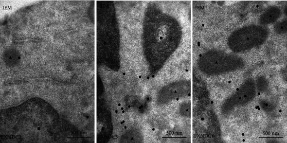
DLBCL is the most common type of non-Hodgkin lymphoma with a substantial group of patients suffering a poor prognosis. Therefore more specific markers are required for better understanding of disease biology and treatment. This study demonstrates that testis-specific antioxidant enzymes TXNDC2, TXNDC3, and TXNDC6 alongside oxidative stress marker 8-OHdG are expressed in both testicular and systemic DLBCL, and their presence or absence has correlations with clinical risk factors such as the number of extranodal effusion, the appearance of B-symptoms, and treatment response. Biopsy samples were collected from 28 systemic and 21 testicular male DLBCL patients. The samples were histostained with TXNDC2, TXNDC3, TXNDC6, and 8-OHdG, then graded by a hematopathologist blinded to clinical data. Immunoelectron microscopy was used as a second method to confirm the reliability of the acquired immunohistochemistry data. The absence of nuclear TXNDC2 expression in testicular DLBCL cells correlated with worse primary treatment response, cytoplasmic TXNDC3 expression in testicular and systemic DLBCL associated with lower frequency of B-symptoms, and TXNDC6 expression in cytoplasm in systemic DLBCL had a clinical significance with higher LD levels suggesting a role in the biological nature of these lymphomas. Overall, TXNDC3 cytoplasmic expression is correlated with a more positive outcome in both testicular and systemic DLBCL, while TXNDC6 cytoplasmic expression is associated with a negative outcome in systemic DLBCL.


Conflict of interest statement

No potential conflict of interest was reported by the authors.

Figures

Figure 1
Figure 1
The expression of 8-OHdG in the patient sample set. Dot plots represent the Histoscore (range 0-300), and micrographs represent the average expression of 8-OHdG in the diagnostic groups. Kruskal-Wallis test was used to calculate the p value. NS: not significant.
Figure 2
Figure 2
The expression of TXNDC2 in the patient sample set and in the cell culture model. Dot plots represent the Histoscore (range 0-300), and micrographs represent the average expression of TXNDC2 in the diagnostic groups. Western blot with three biological replicates of two different DLBCL cell lines shows that TXNDC2 is expressed in these cell lines representing systemic lymphomas. Kruskal-Wallis test was used to calculate the p value. NS: not significant.
Figure 3
Figure 3
The expression of TXNDC3 in the patient sample set and in the cell culture model. Dot plots represent the Histoscore (range 0-300) and micrographs represent the average expression of TXNDC3 in the diagnostic groups. Western blot with three biological replicates of two different DLBCL cell lines shows that TXNDC3 is expressed in these systemic lymphoma cell lines. Kruskal-Wallis test was used to calculate the p value. NS: not significant; H-score: Histoscore.
Figure 4
Figure 4
The expression of TXNDC6 in the patient sample set and in the cell culture model. Box plots represent the Histoscore (range 0-300), and micrographs represent the average expression of TXNDC6 in the diagnostic groups. Western blot with three biological replicates of two different DLBCL cell lines shows that TXNDC6 is expressed in these systemic lymphoma cell lines. Kruskal-Wallis test was used to calculate the p value. NS: not significant; H-score: Histoscore
Figure 5
Figure 5
Immunoelectron microscopy (IEM) showing the localization of the expression of TXNDC2, TXNDC3, and TXNDC6 which are marked as circles. Triangles represent mitochondria and squares nuclei.

Similar articles

Cited by

References

    1. Shaffer A. L., Rosenwald A., Staudt L. M. Lymphoid malignancies: the dark side of B-cell differentiation. Nature Reviews. Immunology. 2002;2(12):920–933. doi: 10.1038/nri953. - DOI - PubMed
    1. Crump M., Neelapu S. S., Farooq U., et al. Outcomes in refractory diffuse large B-cell lymphoma: results from the international SCHOLAR-1 study. Blood. 2017;130(16):1800–1808. doi: 10.1182/blood-2017-03-769620. - DOI - PMC - PubMed
    1. Pui C.-H., Thiel E. Central Nervous System Disease in Hematologic Malignancies: Historical Perspective and Practical Applications. Seminars in Oncology. 2009;36(4, Supplement 2):S2–16. doi: 10.1053/j.seminoncol.2009.05.002. - DOI - PMC - PubMed
    1. Booman M., Szuhai K., Rosenwald A., et al. Genomic alterations and gene expression in primary diffuse large B-cell lymphomas of immune-privileged sites: the importance of apoptosis and immunomodulatory pathways. The Journal of Pathology. 2008;216(2):209–217. doi: 10.1002/path.2399. - DOI - PubMed
    1. Karihtala P., Soini Y. Reactive oxygen species and antioxidant mechanisms in human tissues and their relation to malignancies. APMIS. 2007;115(2):81–103. doi: 10.1111/j.1600-0463.2007.apm_514.x. - DOI - PubMed

MeSH terms