. 2021 Jan 14;12(2):236-241.
doi: 10.1021/acsmedchemlett.0c00605. eCollection 2021 Feb 11.

Development of a Hematopoietic Prostaglandin D Synthase-Degradation Inducer

Affiliations

Development of a Hematopoietic Prostaglandin D Synthase-Degradation Inducer

Hidetomo Yokoo et al. ACS Med Chem Lett. .

Abstract

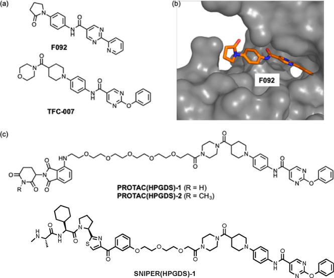
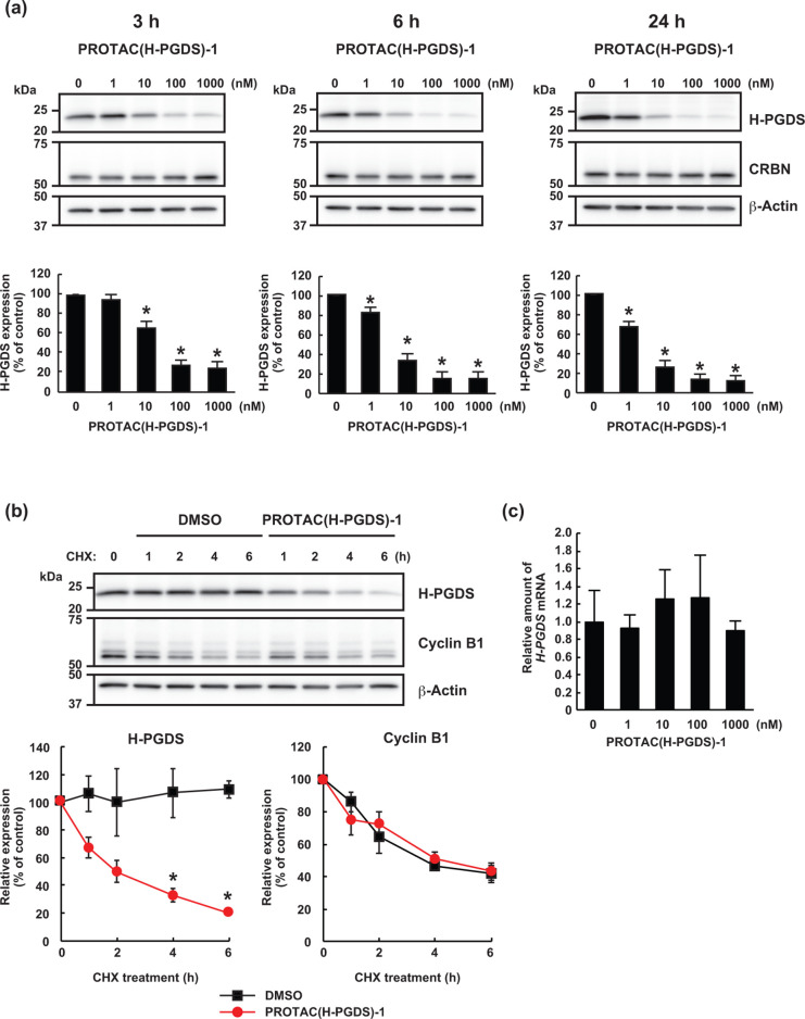
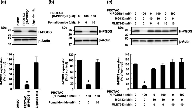
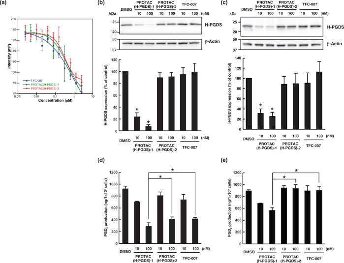
Although hematopoietic prostaglandin D synthase (H-PGDS) is an attractive target for treatment of a variety of diseases, including allergic diseases and Duchenne muscular dystrophy, no H-PGDS inhibitors have yet been approved for treatment of these diseases. Therefore, the development of novel agents having other modes of action to modulate the activity of H-PGDS is required. In this study, a chimeric small molecule that degrades H-PGDS via the ubiquitin-proteasome system, PROTAC(H-PGDS)-1, was developed. PROTAC(H-PGDS)-1 is composed of two ligands, TFC-007 (that binds to H-PGDS) and pomalidomide (that binds to cereblon). PROTAC(H-PGDS)-1 showed potent activity in the degradation of H-PGDS protein via the ubiquitin-proteasome system and in the suppression of prostaglandin D2 (PGD2) production. Notably, PROTAC(H-PGDS)-1 showed sustained suppression of PGD2 production after the drug removal, whereas PGD2 production recovered following removal of TFC-007. Thus, the H-PGDS degrader-PROTAC(H-PGDS)-1-is expected to be useful in biological research and clinical therapies.


Conflict of interest statement

The authors declare no competing financial interest.

Figures

Figure 1
Figure 1
(a) Chemical structures of the H-PGDS inhibitors F092 and TFC-007. (b) X-ray crystal structure of H-PGDS with F092 (PDB: 5YWX). (c) Chemical structures of PROTAC(H-PGDS)-1, PROTAC(H-PGDS)-2, and SNIPER(H-PGDS)-1.
Figure 2
Figure 2
PROTAC(H-PGDS)-1 is a degrader for H-PGDS protein. (a) KU812 cells were incubated with the indicated concentration of PROTAC(H-PGDS)-1 for the indicated time. The H-PGDS/β-actin ratio was normalized by the vehicle control as 100. The data in the bar graph are means ± SD (n = 3). (b) Turnover of H-PGDS protein in KU812 cells after treatment with 10 μg/mL cycloheximide (CHX) in the presence or absence of 100 nM PROTAC(H-PGDS)-1 for the indicated periods. The H-PGDS/β-actin and cyclin B1/β-actin ratios were normalized by the time 0 control as 100. The data in the graphs are means ± SD (n = 3). (c) Expression of H-PGDS mRNA in KU812 cells. Cells were incubated with the indicated concentration of PROTAC(H-PGDS)-1 for 6 h. Expression levels are relative to vehicle treatment, which was arbitrarily set to 1. The data in the bar graph are means ± SD (n = 3). *P < 0.01 compared with vehicle-treated control in a two-tailed Student’s t test.
Figure 3
Figure 3
Involvement of the ubiqutin-proteasome system in the PROTAC(H-PGDS)-1-induced degradation of H-PGDS protein. (a) KU812 cells were incubated with 1 μM PROTAC(H-PGDS)-1 or the ligand mixture (TFC-007 and pomalidomide, 1 μM each) for 6 h. (b) Competition assay using an excess amount of pomalidomide with PROTAC(H-PGDS)-1 in KU812 cells. Cells were incubated with 100 nM PROTAC(H-PGDS)-1 and/or 10 μM pomalidomide for 6 h. (c) Effect of MG132 and MLN7243 on the protein knockdown activity of PROTAC(H-PGDS)-1 in KU812 cells. Cells were incubated with 100 nM PROTAC(H-PGDS)-1 in the presence or absence of 10 μM MG132 or 10 μM MLN7243 for 6 h. The H-PGDS/β-actin ratio was normalized by the vehicle control as 100. The data in the bar graphs are means ± SD (n = 3). *P < 0.01 compared to vehicle-treated control in a two-tailed Student’s t test.
Figure 4
Figure 4
Comparison of the degradation and inhibition of H-PGDS. (a) Fluorescence polarization assays of the binding affinity between H-PGDS and TFC-007, PROTAC(H-PGDS)-1, and PROTAC(H-PGDS)-2. (b, c) KU812 cells were incubated with the indicated concentration of the compounds for 24 h (b) and then washed four times to remove the compounds and incubated in compound-free medium for 6 h (c). The H-PGDS/β-actin ratio was normalized by the vehicle control as 100. The data in the bar graph are means ± SD (n = 3). *P < 0.01 compared to vehicle-treated control in a two-tailed Student’s t test. (d, e) KU812 cells were incubated with the indicated concentration of the compounds for 24 h. Then, the cells were incubated with 5 μM A23187 in the presence of each compound for 30 min (d), or the cells were washed four times to remove the compounds, incubated in compound-free medium for 6 h, and incubated with 5 μM A23187 for 30 min (e). PGD2 levels in the medium were measured. The data in the bar graph are means ± SD (n = 3). *P < 0.05 in a two-tailed Student’s t test.

References

    1. Lewis R. A.; Soter N. A.; Diamond P. T.; Austen K. F.; Oates J. A.; Roberts L. J. Prostaglandin D2 Generation after Activation of Rat and Human Mast Cells with Anti-IgE. J. Immunol. 1982, 129, 1627–1631. - PubMed
    1. Matsuoka T.; Hirata M.; Tanaka H.; Takahashi Y.; Murata T.; Kabashima K.; Sugimoto Y.; Kobayashi T.; Ushikubi F.; Aze Y.; Eguchi N.; Urade Y.; Yoshida N.; Kimura K.; Mizoguchi A.; Honda Y.; Nagai H.; Narumiya S. Prostaglandin D2 as a Mediator of Allergic Asthma. Science 2000, 287, 2013–2017. 10.1126/science.287.5460.2013. - DOI - PubMed
    1. Urade Y.; Hayaishi O. Biochemical, Structural, Genetic, Pysiological, and Pathophysiological Features of Lipocalin-type Prostaglandin D Synthase. Biochim. Biophys. Acta, Protein Struct. Mol. Enzymol. 2000, 1482, 259–271. 10.1016/S0167-4838(00)00161-8. - DOI - PubMed
    1. Takeshita E.; Komaki H.; Shimizu-Motohashi Y.; Ishiyama A.; Sasaki M.; Takeda S. A Phase I Study of TAS-205 in Patients with Duchenne Muscular Dystrophy. Ann. Clin. Transl. Neurol. 2018, 5, 1338–1349. 10.1002/acn3.651. - DOI - PMC - PubMed
    1. Rittchen S.; Heinemann A. Therapeutic Potential of Hematopoietic Prostaglandin D2 Synthase in Allergic Inflammation. Cells 2019, 8, 619.10.3390/cells8060619. - DOI - PMC - PubMed