. 2019 Dec;6(12):125410.
doi: 10.1088/2053-1591/ab597a. Epub 2019 Nov 29.

Non-invasive acoustic fabrication methods to enhance collagen hydrogel bioactivity

Affiliations

Non-invasive acoustic fabrication methods to enhance collagen hydrogel bioactivity

Emma G Norris et al. Mater Res Express. 2019 Dec.

Abstract

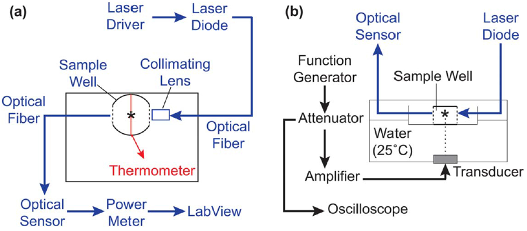
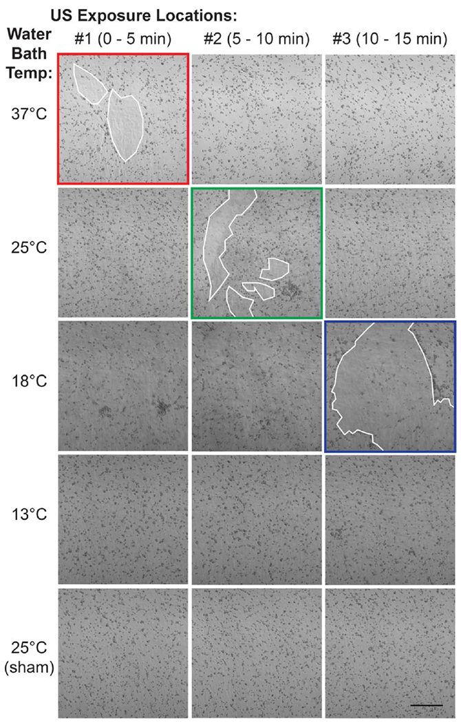
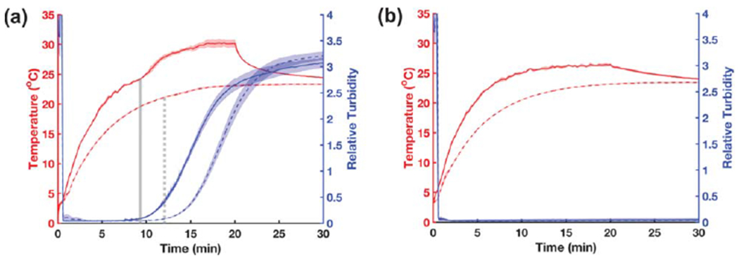
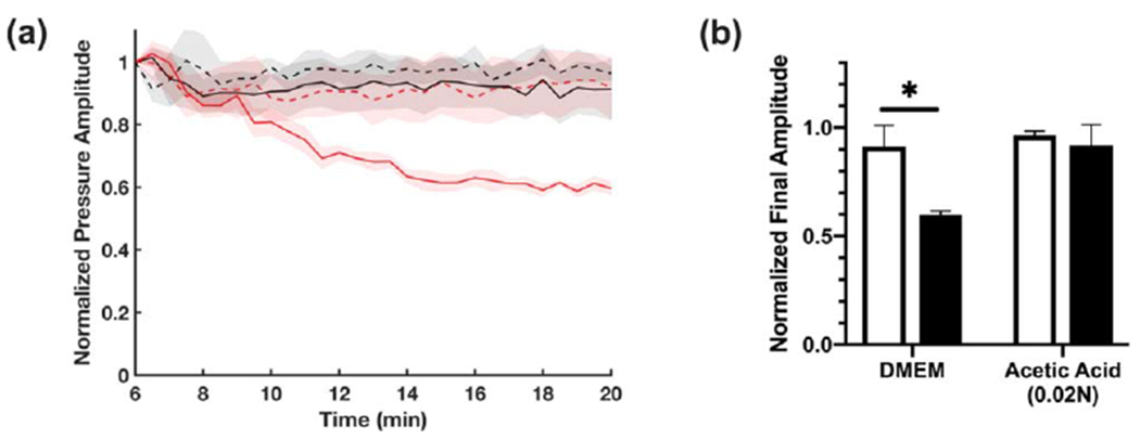
Much attention has focused recently on utilizing components of the extracellular matrix (ECM) as natural building blocks for a variety of tissue engineering applications and regenerative medicine therapies. Consequently, new fabrication methods are being sought to enable molecular control over the structural characteristics of ECM molecules in order to improve their biological function. Exposing soluble collagen to acoustic forces associated with ultrasound propagation produces localized variations in collagen microfiber organization that in turn, promote cell behaviors essential for tissue regeneration, including cell migration and matrix remodeling. In the present study, mechanisms by which ultrasound interacts with polymerizing collagen to produce functional changes in collagen microstructure were investigated. The rate of collagen polymerization was manipulated by adjusting the pH of collagen solutions and the temperature at which gels were polymerized. Results demonstrate that the phase transition of type I collagen from fluid to gel triggered a simultaneous increase in acoustic absorption. This phase transition of collagen involves the lateral growth of early-stage collagen microfibrils and importantly, corresponded to a defined period of time during which exposure to ultrasound introduced both structural and functional changes to the resultant collagen hydrogels. Together, these experiments isolated a critical window in the collagen fiber assembly process during which mechanical forces associated with ultrasound propagation are effective in producing structural changes that underlie the ability of acoustically-modified collagen hydrogels to stimulate cell migration. These results demonstrate that changes in material properties associated with collagen polymerization are a fundamental component of the mechanism by which acoustic forces modify collagen biomaterials to enhance biological function.

Keywords: acoustics; biofabrication; collagen; tissue engineering; ultrasound.


Conflict of interest statement

Declarations of interest none

Figures

Figure 1.
Figure 1.
Schematic of optical monitoring system for ultrasound-exposed collagen (a) Top-down view of ultrasound exposure system illustrating components of the optical and temperature monitoring systems. Parallel sides of the plastic well were cut out and replaced with glass windows (dashed lines). Light from a 405-nm laser diode was directed into the collagen sample via a collimating lens and received on the opposite side by an optical sensor. Optical power measurements were acquired by LabView software. A type-T wire thermocouple (red line) was placed below the optical beam path and the temperature of the sample monitored by a digital thermometer. The spectroscopy plate was placed in an ultrasound exposure chamber such that the thermocouple junction and light paths coincided with the 3-mm ultrasound beam (asterisk), (b) Ultrasound fields (8.8 MHz, 7.9 W cm−2) were generated using an unfocused piezoceramic transducer mounted at the bottom of a temperature-controlled water bath. The plate was allowed to equilibrate to water bath temperature prior to sample addition. Black, ultrasound exposure system; blue, optical monitoring system; red, temperature monitoring system.
Figure 2.
Figure 2.
Effect of collagen polymerization on ultrasound-induced heating (a) Representative image illustrating the cellular response to acoustically-modified collagen hydrogels. FN-null MEFs (4.7 × 10⁴ cells cm−2) were seeded on the surface of collagen gels polymerized in the presence of 8.8-MHz ultrasound for 15 min (7.9 W cm−2). Following 24 h of culture, cells on the surface of acoustically-modified collagen gels migrated into large linear aggregates (white arrows), leaving behind cell-free areas (white asterisks). Black asterisk indicates the center of the acoustic beam. Scale bar = 500 μm. (b) The temperature of collagen (0.8 mg ml−1) was measured at the center of the acoustic beam during exposure to 7.8-MHz ultrasound at an intensity of 0 (sham, open circles) or 10 W cm−2 (filled circles). Collagen solutions were prepared in DMEM and the pH adjusted to either 6.1 (gray), or 7.4 (black) with 0.02 N acetic acid. All exposures were conducted in a chamber with the water temperature set to 25 °C. Temperature was recorded every 15 s for the duration of the exposure. Data are presented as mean ± SEM for n ⩾ 3 replicates per condition.
Figure 3.
Figure 3.
Aliquots of neutralized soluble collagen (0.8 mg ml−1) were transferred to exposure plates positioned in a 25 °C water bath. Collagen polymerization was initiated, and samples were exposed to ultrasound (8.8 MHz, 7.9 W cm−2) for 5 min during 1 of 3 non-overlapping time periods spanning the polymerization process: 0–5, 5–10, or 10–15 min. Control collagen samples were sham-exposed. Polymerized gels were imaged using SHG microscopy. Maximum intensity projections were obtained through a depth of 100 μm and assembled using FIJI software. Images represent 1 of at least 3 experiments from gels fabricated on independent days. Scale bar = 100 μm.
Figure 4.
Figure 4.
Onset of second stage of ultrasound-induced heating depends on collagen polymerization rate (a) The temperature of neutralized collagen solutions (0.8 mg mL−1 in DMEM) was measured at the center of the acoustic beam during exposure to 8.8-MHz ultrasound (7.9 W cm−2). The polymerization rate of collagen was adjusted by setting the water bath temperature to 13 (black), 18 (blue), 25 (green), or 37 °C (red). Temperature was recorded every 15 s for the duration of the exposure. Data are presented as mean ± SEM for n ⩾ 3 replicates per condition. (b) Schematic illustrating multipoint ultrasound exposures trategy. Collagen solutions were exposed to 8.8-MHz ultrasound (7.9 W cm−2) for 5 min at each of 3 positions (asterisks) over the course of 15 min. The half-maximal beam width of the acoustic field was 3 mm (dashed circles). The center of each exposure location (asterisks) was separated by step sizes of 5 mm to ensure no overlap between exposure locations. Scale = 1 mm/division.
Figure 5.
Figure 5.
Pro-migratory activity is introduced during second stage of ultrasound-induced heating. Aliquots of neutralized soluble collagen (0.8 mg ml−1) were exposed to 8.8-MHz ultrasound (7.9 W cm−2) or sham-exposed at 3 different, sequential locations for 5 min each, for a total exposure of l5 min. Additionally, the rate of collagen polymerization was varied by setting the water bath temperature to 37, 25, 18, or 13 °C, so that the second stage of ultrasound-induced heating coincided with the first (37 °C, red), second (25 °C, green), or third (18 °C, blue) exposure location. Collagen polymerization did not occur within the 15-min exposure period when the temperature was set to 13 °C. All samples were allowed to polymerize fully post-exposure before FN-null MEFs (4.7 × 10⁴ cells cm−2) were seeded on gel surfaces. After 24 h, phase contrast images were captured at each of the 3 exposure locations. White outlines indicate cell-free areas on the collagen gel surface. Images represent 1 of at least 3 independent experiments. Scale bar = 500 μm.
Figure 6.
Figure 6.
Turbidity monitoring of collagen polymerization during ultrasound exposure Aliquots of neutralized collagen (a) or DMEM alone (b) were either exposed to 8.8-MHz ultrasound (solid lines, 7.9 W cm−2) or sham-exposed (dashed lines, 0 W cm−2) for 20 min in a 25 °C water bath; data were collected for an additional 10 min after the sound was turned off. The optical power (405 nm) transmitted across a 3.7-cm path length was recorded at a 10-Hz sampling rate. Data are presented as optical turbidity (blue) relative to transmission through an empty well. Temperature (red) at the center of each sample was simultaneously recorded every 15 s using a type-T wire thermocouple. In panel (a), vertical gray lines indicate the end of the optical lag phase for ultrasound- (solid) and sham-exposed (dashed) samples. Data are presented as mean ± SEM for n = 3 replicates per condition.
Figure 7.
Figure 7.
Ultrasound does not alter the acoustic absorption coefficient of collagen directly. Collagen samples (0.8 mg ml−1) with embedded thermocouples were first polymerized in the presence or absence of 7.8-MHz ultrasound (10 W cm−2). Fully polymerized samples were then allowed to equilibrate to water bath temperature. Both non-exposed (open circles) and ultrasound-exposed (filled circles) collagen gels were subsequently heated with 7.8-MHz ultrasound (10 W cm−2) and the collagen temperature within the acoustic beam was recorded every 5 s for 3 min (a). (b) The ultrasound-induced heating rate was calculated for the initial 15 s of each recording. No differences were observed in the ultrasound-induced heating rate for collagen polymerized under sham (white), or ultrasound conditions (black).
Figure 8.
Figure 8.
Collagen fiber assembly increases the acoustic absorption coefficient. Aliquots of soluble collagen (1.0 mg ml−1 were transferred into the wells of modified tissue culture plates with a wire thermocouple placed at the center. Samples were allowed to equilibrate for 1 h in the absence of ultrasound. During this time, collagen polymerization either proceeded (neutralized DMEM, (a), or was prevented by maintaining acidic conditions (0.02 N acetic acid in PBS, (b). The temperature at the center of each sample was recorded during subsequent exposure to 8.8-MHz ultrasound at intensities of 1.7 (black), 3.6 (blue), and 7.9 W cm−2 (red). Each graph represents a set of recordings from 1 of at least 3 independent samples. (c) The initial ultrasound-induced heating rate was calculated for polymerized (black) and non-polymerized (white) samples by performing a linear regression over the first 10 s of ultrasound exposure. Data are presented as mean ± SEM for n ⩾ 4 recordings from at least 3 independent samples per condition. Significantly different means, *P < 0.05 by t-test with Holm-Sidak’s post-hoc test for multiple comparisons.
Figure 9.
Figure 9.
Effect of collagen polymerization on acoustic attenuation (a) Aliquots of soluble collagen solution (2.0 mg ml−1, solid lines) or vehicle control (dashed lines) were prepared under either neutralized (red, DMEM) or acidic (black, 0.02 N acetic acid in PBS) conditions. Cold samples were placed in a 4-cm long cylindrical sample holder with Saran membranes on either side. Acoustic attenuation measurements were obtained by a pulse-echo technique using a 28 ns ultrasound pulse (38 MHz center frequency) reflected by a steel disk placed at the bottom of the propagation path. The amplitude of the signal received from the reflector was recorded every 30 s and plotted beginning 6 min after sample addition (e.g., prior to collagen polymerization), as the normalized mean ± SEM for n ⩾ 3 replicates per condition. (b) Echo amplitude at 20 min normalized to initial amplitude (6 min) for vehicle control (white), or 2.0 mg ml−1 collagen (black) samples. Data are presented as mean ± SEM for n ⩾ 3 replicates per condition. Significantly different means, *P < 0.05 by t-test with Holm-Sidak’s post-hoc test for multiple comparisons.

Similar articles

Cited by

References

    1. Langer R and Vacanti JP 1993. Tissue engineering Science 260 920–6 - PubMed
    1. Abou Neel EA, Bozec L, Knowles JC, Syed O, Mudera V, Day R and Keun Hyun J 2013. Collagen—emerging collagen based therapies hit the patient Adv. Drug Delivery Rev 65 429–56 - PubMed
    1. Kadler KE, Baldock C, Bella JandBoot-Handford RP 2007. Collagens at a glance J. Cell Sci 120 1955–8 - PubMed
    1. Cen L, Liu W, Cui L, Zhang W and Cao Y 2008. Collagen tissue engineering: development of novel biomaterials and applications Pediatr Res 63 492–6 - PubMed
    1. Schleifenbaum S. et al. A cellularization-induced changes in tensile properties are organ specific—an in-vitro mechanical and structural analysis of porcine soft tissues. PLoS One. 2016;11:e0151223. - PMC - PubMed

LinkOut - more resources