

Photoacoustic Imaging of Tattoo Inks: Phantom and Clinical Evaluation
- PMID: 33604062
- PMCID: PMC7889065
- DOI: 10.3390/app10031024
Photoacoustic Imaging of Tattoo Inks: Phantom and Clinical Evaluation
Abstract
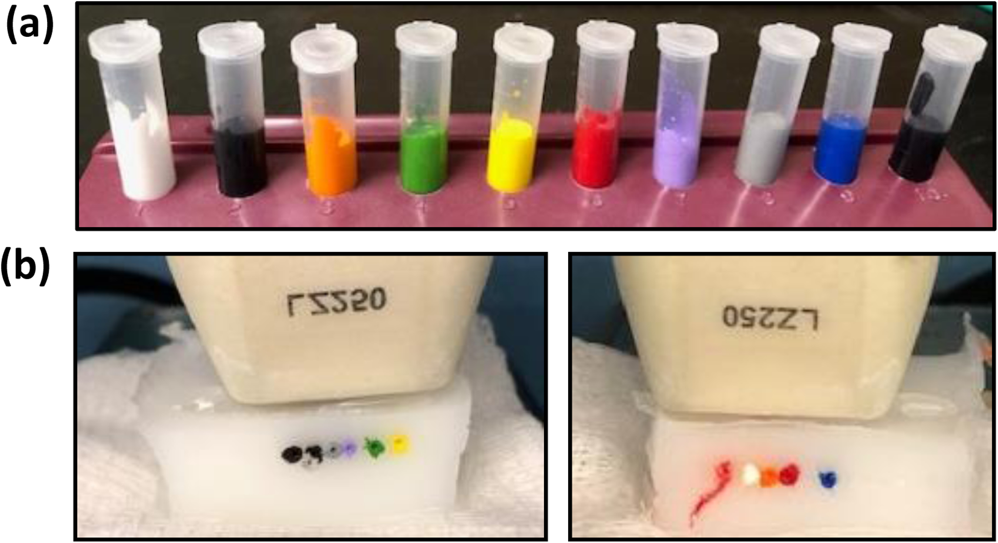
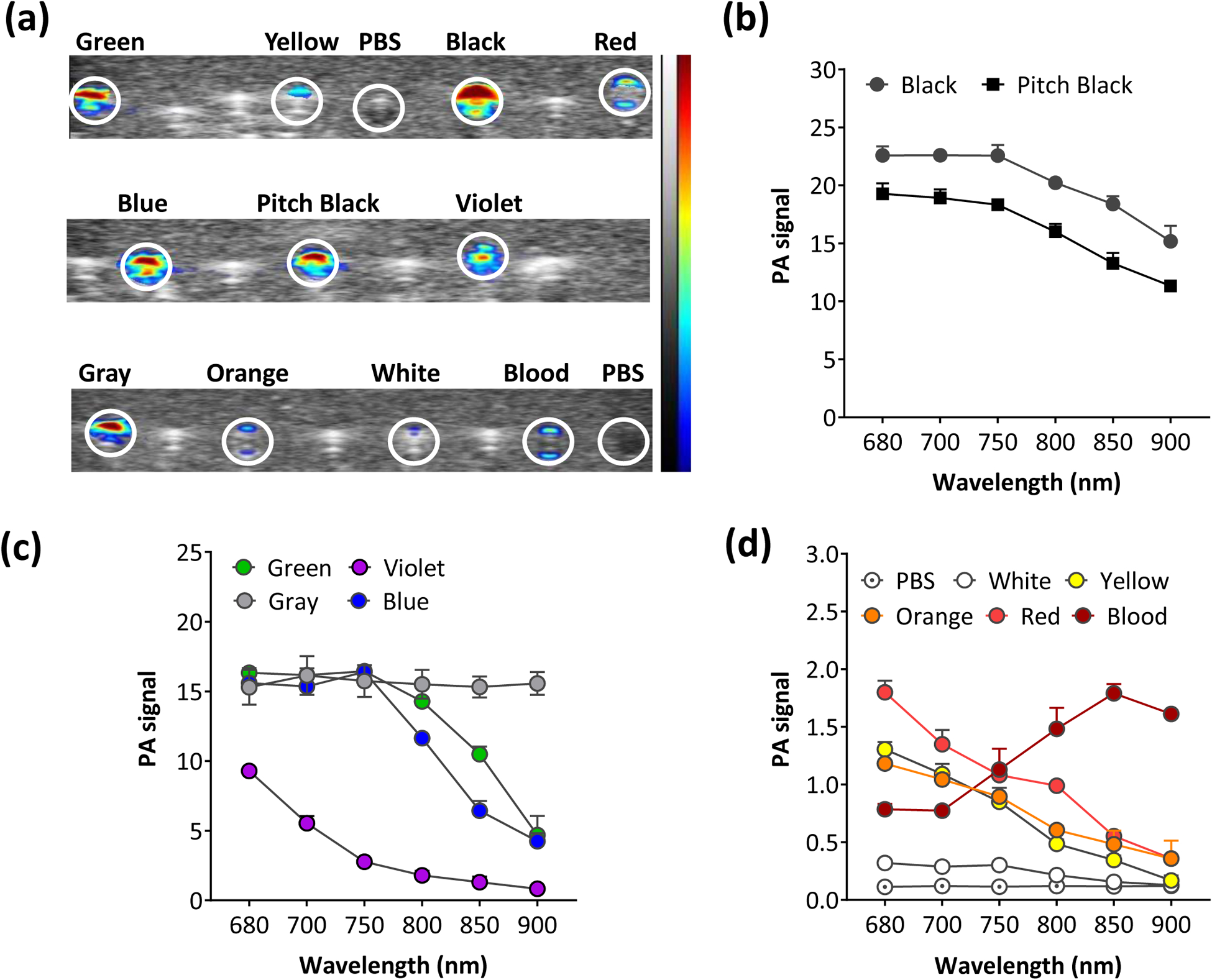
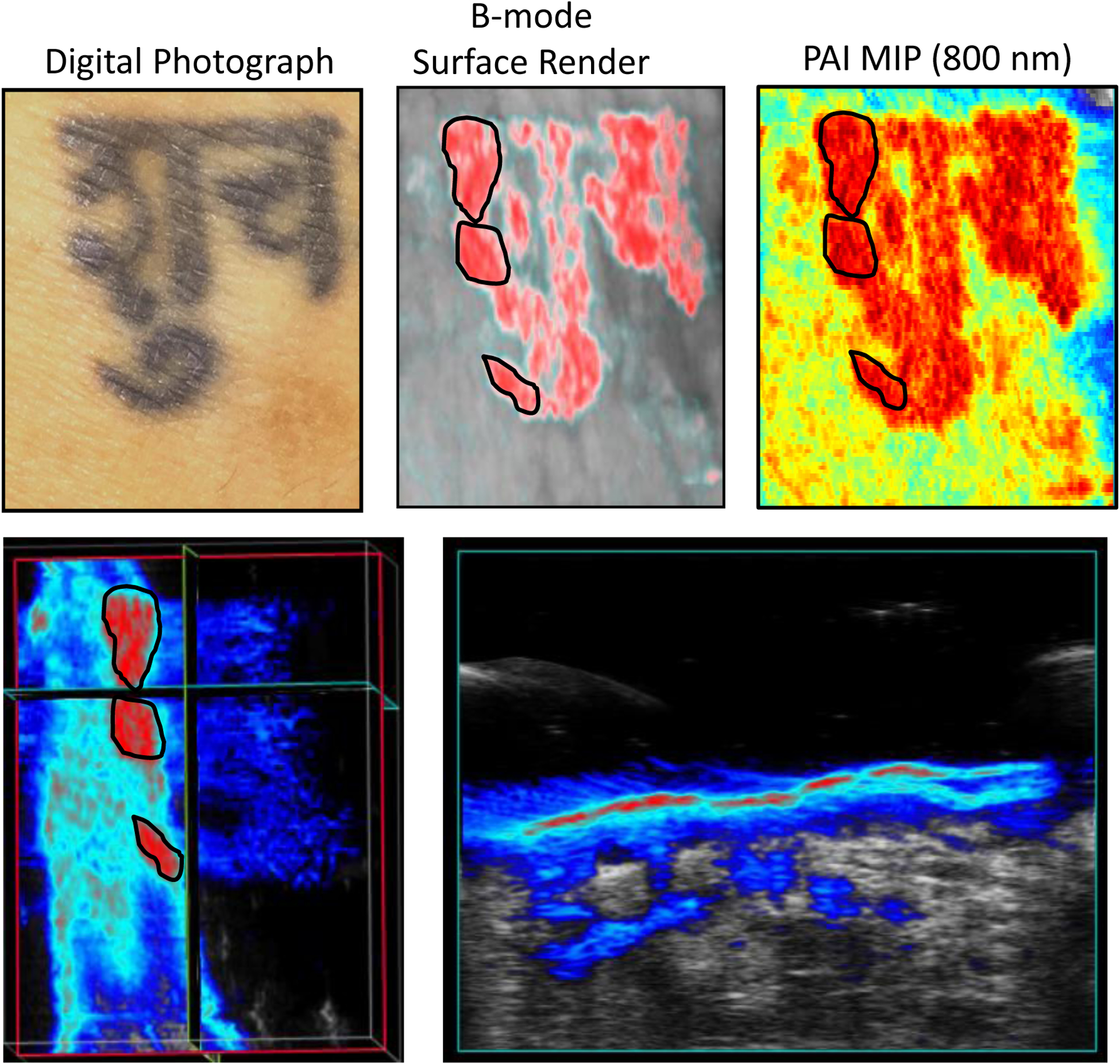
Photoacoustic imaging (PAI) is a novel hybrid imaging modality that provides excellent optical contrast with the spatial resolution of ultrasound in vivo. The method is widely being investigated in the clinical setting for diagnostic applications in dermatology. In this report, we illustrate the utility of PAI as a non-invasive tool for imaging tattoos. Ten different samples of commercially available tattoo inks were examined for their optoacoustic properties in vitro. In vivo PAI of an intradermal tattoo on the wrist was performed in a healthy human volunteer. Black/gray, green, violet and blue colored pigments provided higher levels of PA signal compared to white, orange, red and yellow pigments in vitro. PAI provided excellent contrast and enabled accurate delineation of the extent of the tattoo in the dermis. Our results reveal the photoacoustic properties of tattoo inks and demonstrate the potential clinical utility of PAI for intradermal imaging of tattoos. PAI may be useful as a clinical adjunct for objective preoperative evaluation of tattoos and potentially to guide/monitor laser-based tattoo removal procedures.
Keywords: dermatology; photoacoustic imaging; tattoo; ultrasound.
Conflict of interest statement
Conflicts of Interest: The authors declare no conflict of interest.
Figures
Similar articles
-
In vitro and in vivo laser treatments of tattoos: high efficiency and low fluences.Arch Dermatol. 2010 Jan;146(1):39-45. doi: 10.1001/archdermatol.2009.321. Arch Dermatol. 2010. PMID: 20083691
-
Laser removal of tattoos.Am J Clin Dermatol. 2001;2(1):21-5. doi: 10.2165/00128071-200102010-00004. Am J Clin Dermatol. 2001. PMID: 11702617 Review.
-
Colored Tattoo Ink Screening Method with Optical Tissue Phantoms and Raman Spectroscopy.Materials (Basel). 2021 Jun 8;14(12):3147. doi: 10.3390/ma14123147. Materials (Basel). 2021. PMID: 34201157 Free PMC article.
-
A novel titanium sapphire picosecond-domain laser safely and effectively removes purple, blue, and green tattoo inks.Lasers Surg Med. 2018 May 20;50(7):704-10. doi: 10.1002/lsm.22942. Online ahead of print. Lasers Surg Med. 2018. PMID: 29781161 Free PMC article.
-
Lasers in tattoo and pigmentation control: role of the PicoSure(®) laser system.Med Devices (Auckl). 2016 May 2;9:63-7. doi: 10.2147/MDER.S77993. eCollection 2016. Med Devices (Auckl). 2016. PMID: 27194919 Free PMC article. Review.
References
-
- Hamblin MR, Avci P, Gupta GK, editors. Imaging in dermatology. Academic Press; 2016.
-
- Hibler BP, Qi Q, Rossi AM. Current state of imaging in dermatology. Semin Cutan Med Surg. 2016; 35:2–8. - PubMed
-
- Dubois A, Levecq O, Azimani H, et al. Line-field confocal optical coherence tomography for high-resolution noninvasive imaging of skin tumors. J Biomed Opt 2018; 23: 106007. - PubMed
Grants and funding
LinkOut - more resources
Full Text Sources