Case Reports
. 2021 Jan 28:2021:5952724.
doi: 10.1155/2021/5952724. eCollection 2021.

Rare Case of Spinal Neurosarcoidosis with Concomitant Epidural Lipomatosis

Affiliations
Case Reports

Rare Case of Spinal Neurosarcoidosis with Concomitant Epidural Lipomatosis

Nesreen Jaafar et al. Case Rep Neurol Med. .

Abstract

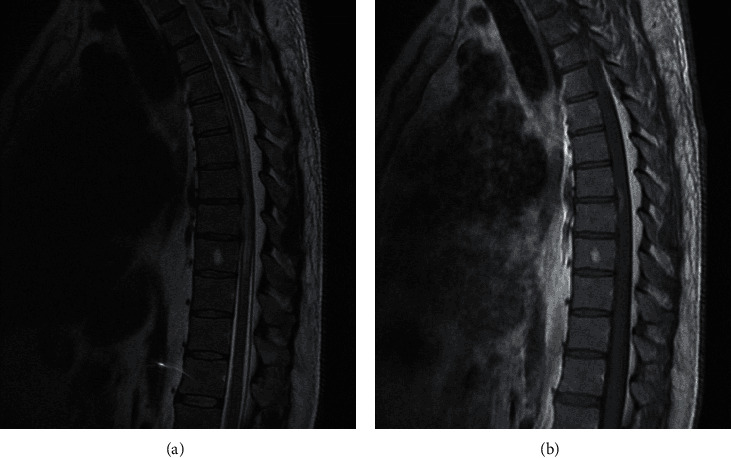
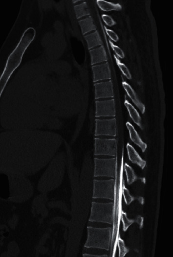
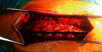
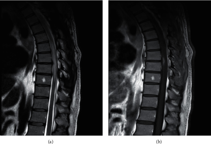
Introduction: Spinal neurosarcoidosis is a rare disease that can manifest as myelopathy, radiculopathy, or cauda equine syndrome. Spinal epidural lipomatosis is also a rare condition resulting from overgrowth of epidural fat tissue causing compressive myelopathy. To our knowledge, there are no reports linking epidural lipomatosis and spinal neurosarcoidosis. Case Report. We describe a case of progressive myelitis in the presence of concomitant spinal neurosarcoidosis and epidural lipomatosis which was a challenging diagnosis with complete response to treatment after addressing both diseases. Both etiologies are inflammatory in nature and share similar expression of inflammatory factors such as TNF-α and IL-1β.

Conclusion: The common inflammatory process involved in these two diseases might explain a pathophysiological interconnection between both diseases that may underlie their concomitant development in our patient. If these two diseases are interconnected, in their pathophysiological mechanism remains a hypothesis that will need further investigation.


Conflict of interest statement

The authors declare that they have no conflicts of interest.

Figures

Figure 1
Figure 1
(a) Sagittal T2 MRI of dorsal spine showing high T2 signal with cord edema from T4 to T9 levels with thickened epidural fat tissue posteriorly. (b) Sagittal T1 MRI with gadolinium of dorsal spine showing an enhancing lesion at T7-T8 level.
Figure 2
Figure 2
CT myelogram showing obstruction of CSF flow extending from T4 to T9 vertebrae.
Figure 3
Figure 3
Intraoperative image showing the thickened epidural fat before excision.
Figure 4
Figure 4
(a) Sagittal T2 MRI showing resolution of the epidural lipomatosis with resolution of the mass effect on the spinal cord and decrease in the extent of the lesion (from T6 to T9). (b) Sagittal T1 MRI with gadolinium showing mild decrease in the size of enhancing lesion.
Figure 5
Figure 5
CT chest showing bilateral mediastinal and hilar adenopathy.
Figure 6
Figure 6
(a) Sagittal T2 MRI of whole spine after 2 months of treatment with mycophenolate mofetil showing decrease in T2 signal involving T7-T8. (b) Sagittal T1 MRI with gadolinium after 2 months of treatment showing further decrease in extent of enhancing lesion.
Figure 7
Figure 7
Sagittal T1 MRI with gadolinium showing the decrease in extent of enhancing lesion from initial MRI, after surgery, and after treatment.

Similar articles

Cited by

  • Spinal Epidural Lipomatosis: A Comprehensive Review.
    Walker PB, Sark C, Brennan G, Smith T, Sherman WF, Kaye AD. Walker PB, et al. Orthop Rev (Pavia). 2021 Jul 11;13(2):25571. doi: 10.52965/001c.25571. eCollection 2021. Orthop Rev (Pavia). 2021. PMID: 34745483 Free PMC article. Review.
  • [Clinical research progress of spinal epidural lipomatosis].
    Zhang L, Xu Y, Fan P, Li X, Wang J, Tao Y, Li X, Wang Y. Zhang L, et al. Zhongguo Xiu Fu Chong Jian Wai Ke Za Zhi. 2023 Oct 15;37(10):1284-1291. doi: 10.7507/1002-1892.202305071. Zhongguo Xiu Fu Chong Jian Wai Ke Za Zhi. 2023. PMID: 37848326 Free PMC article. Review. Chinese.
  • Outcome of posterior decompression for spinal epidural lipomatosis.
    Schmutzer-Sondergeld M, Zimmermann H, Trabold R, Liebig T, Schichor C, Siller S. Schmutzer-Sondergeld M, et al. Acta Neurochir (Wien). 2023 Nov;165(11):3479-3491. doi: 10.1007/s00701-023-05814-0. Epub 2023 Sep 25. Acta Neurochir (Wien). 2023. PMID: 37743433 Free PMC article.

References

    1. Iannuzzi M. C., Rybicki B. A., Teirstein A. S. Sarcoidosis. New England Journal of Medicine. 2007;357(21):2153–2165. doi: 10.1056/nejmra071714. - DOI - PubMed
    1. Stern B. J., Krumholz A., Johns C., Scott P., Nissim J. Sarcoidosis and its neurological manifestations. Archives of Neurology. 1985;42(9):909–917. doi: 10.1001/archneur.1985.04060080095022. - DOI - PubMed
    1. Bradley D. A., Lower E. E., Baughman R. P. Diagnosis and management of spinal cord sarcoidosis. Sarcoidosis, Vasculitis, and Diffuse Lung Diseases: Official Journal of WASOG. 2006;23(1):58–65. - PubMed
    1. Scott A. M., Yinh J., McAlindon T., Kalish R. Two cases of sarcoidosis presenting as longitudinally extensive transverse myelitis. Clinical Rheumatology. 2018;37(10):2899–2905. doi: 10.1007/s10067-018-4144-9. - DOI - PubMed
    1. Wang L., Li Y. Longitudinal ultra-extensive transverse myelitis as a manifestation of neurosarcoidosis. Journal of the Neurological Sciences. 2015;355(1-2):64–67. doi: 10.1016/j.jns.2015.05.017. - DOI - PubMed

Publication types

LinkOut - more resources