

Induction of Functional Hypothalamus and Pituitary Tissues From Pluripotent Stem Cells for Regenerative Medicine
- PMID: 33604493
- PMCID: PMC7880040
- DOI: 10.1210/jendso/bvaa188
Induction of Functional Hypothalamus and Pituitary Tissues From Pluripotent Stem Cells for Regenerative Medicine
Abstract
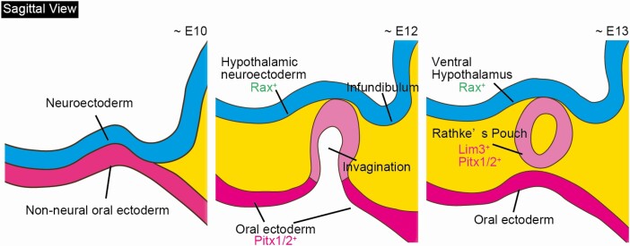
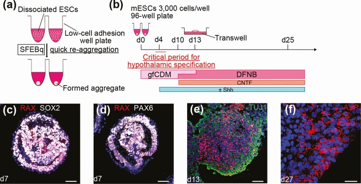
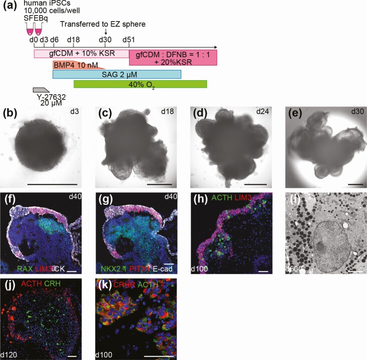
The hypothalamus and pituitary have been identified to play essential roles in maintaining homeostasis. Various diseases can disrupt the functions of these systems, which can often result in serious lifelong symptoms. The current treatment for hypopituitarism involves hormone replacement therapy. However, exogenous drug administration cannot mimic the physiological changes that are a result of hormone requirements. Therefore, patients are at a high risk of severe hormone deficiency, including adrenal crisis. Pluripotent stem cells (PSCs) self-proliferate and differentiate into all types of cells. The generation of endocrine tissues from PSCs has been considered as another new treatment for hypopituitarism. Our colleagues established a 3-dimensional (3D) culture method for embryonic stem cells (ESCs). In this culture, the ESC-derived aggregates exhibit self-organization and spontaneous formation of highly ordered patterning. Recent results have shown that strict removal of exogenous patterning factors during early differentiation efficiently induces rostral hypothalamic progenitors from mouse ESCs. These hypothalamic progenitors generate vasopressinergic neurons, which release neuropeptides upon exogenous stimulation. Subsequently, we reported adenohypophysis tissue self-formation in 3D cultures of mouse ESCs. The ESCs were found to differentiate into both nonneural oral ectoderm and hypothalamic neuroectoderm in adjacent layers. Interactions between the 2 tissues appear to be critically important for in vitro induction of a Rathke's pouch-like developing embryo. Various endocrine cells were differentiated from nonneural ectoderm. The induced corticotrophs efficiently secreted adrenocorticotropic hormone when engrafted in vivo, which rescued hypopituitary hosts. For future regenerative medicine, generation of hypothalamic and pituitary tissues from human PSCs is necessary. We and other groups succeeded in establishing a differentiation method with the use of human PSCs. Researchers could use these methods for models of human diseases to elucidate disease pathology or screen potential therapeutics.
Keywords: differentiation; embryonic stem cells; hypothalamus; induced pluripotent stem cells; pituitary; regenerative medicine.
© The Author(s) 2020. Published by Oxford University Press on behalf of the Endocrine Society.
Figures
References
-
- Hahner S, Spinnler C, Fassnacht M, et al. High incidence of adrenal crisis in educated patients with chronic adrenal insufficiency: a prospective study. J Clin Endocrinol Metab. 2015;100(2):407-416. - PubMed
-
- Zhu X, Gleiberman AS, Rosenfeld MG. Molecular physiology of pituitary development: signaling and transcriptional networks. Physiol Rev. 2007;87(3):933-963. - PubMed
-
- Takuma N, Sheng HZ, Furuta Y, et al. Formation of Rathke’s pouch requires dual induction from the diencephalon. Development. 1998;125(23):4835-4840. - PubMed
-
- Treier M, O’Connell S, Gleiberman A, et al. Hedgehog signaling is required for pituitary gland development. Development. 2001;128(3):377-386. - PubMed
Publication types
LinkOut - more resources
Full Text Sources