

Diabetic Kidney Disease, Endothelial Damage, and Podocyte-Endothelial Crosstalk
- PMID: 33604542
- PMCID: PMC7873832
- DOI: 10.1016/j.xkme.2020.10.005
Diabetic Kidney Disease, Endothelial Damage, and Podocyte-Endothelial Crosstalk
Abstract
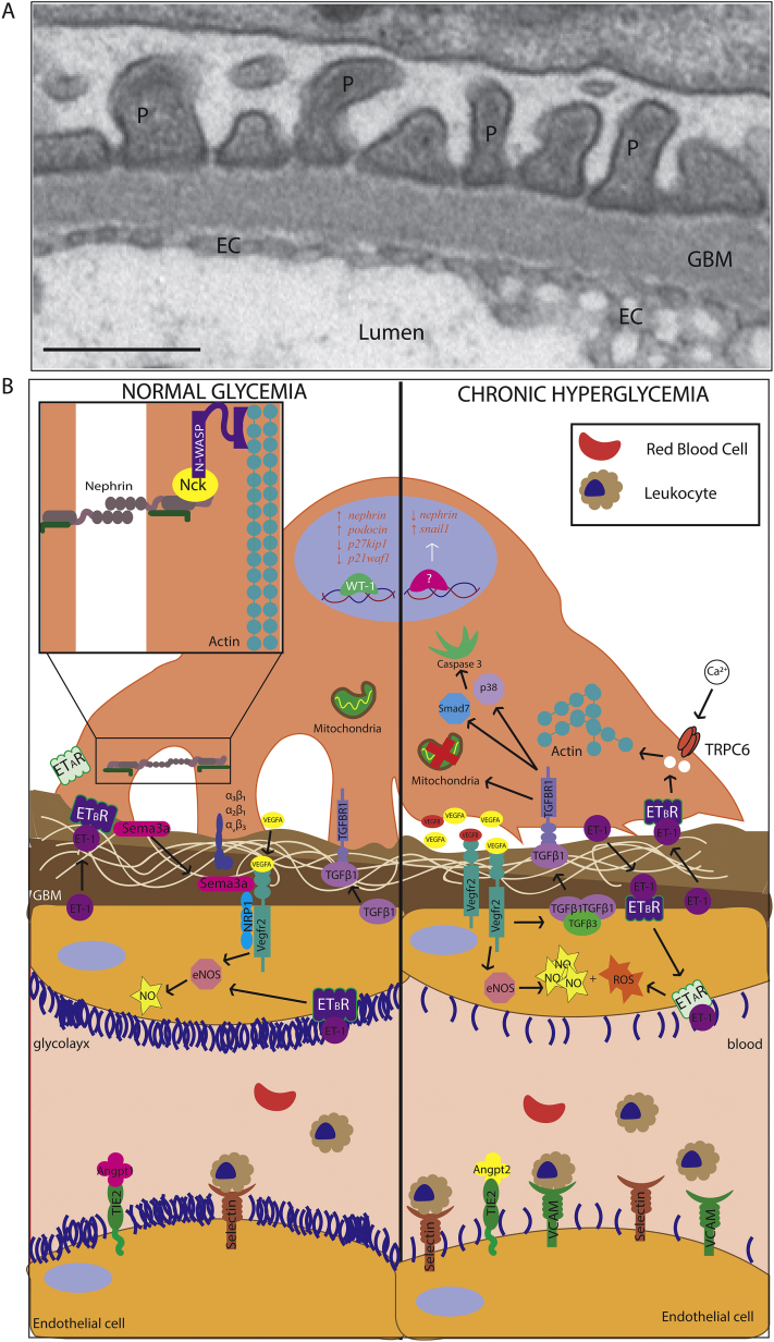
Diabetes-related complications are a significant source of morbidity and mortality worldwide. Diabetic kidney disease is a frequent microvascular complication and a primary cause of kidney failure in patients with diabetes. The glomerular filtration barrier is composed of 3 layers: the endothelium, glomerular basement membrane, and podocytes. Podocytes and the endothelium communicate through molecular crosstalk to maintain filtration at the glomerular filtration barrier. Chronic hyperglycemia affects all 3 layers of the glomerular filtration barrier, as well as the molecular crosstalk that occurs between the 2 cellular layers. One of the earliest events following chronic hyperglycemia is endothelial cell dysfunction. Early endothelial damage is associated with progression of diabetic kidney disease. However, current therapies are based in controlling glycemia and arterial blood pressure without targeting endothelial dysfunction. Disruption of the endothelial cell layer also alters the molecular crosstalk that occurs between the endothelium and podocytes. This review discusses both the physiologic and pathologic communication that occurs at the glomerular filtration barrier. It examines how these signaling components contribute to podocyte foot effacement, podocyte detachment, and the progression of diabetic kidney disease.
Keywords: Podocyte-endothelial crosstalk; albuminuria; diabetic kidney disease; diabetic nephropathy; microvascular.
© 2020 The Authors.
Figures
References
-
- Sena C.M., Pereira A.M., Seiça R. Endothelial dysfunction — a major mediator of diabetic vascular disease. Biochim Biophys Acta. 2013;1832:2216–2231. - PubMed
Publication types
LinkOut - more resources
Full Text Sources
Other Literature Sources
