

Macrophages as host, effector and immunoregulatory cells in leishmaniasis: Impact of tissue micro-environment and metabolism
- PMID: 33604563
- PMCID: PMC7885870
- DOI: 10.1016/j.cytox.2020.100041
Macrophages as host, effector and immunoregulatory cells in leishmaniasis: Impact of tissue micro-environment and metabolism
Abstract
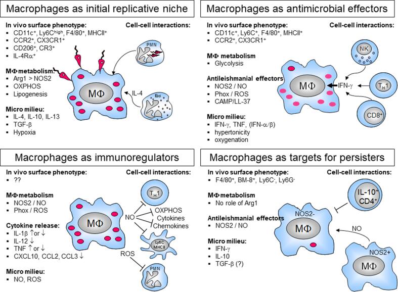
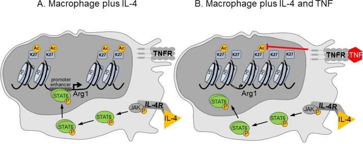
Leishmania are protozoan parasites that predominantly reside in myeloid cells within their mammalian hosts. Monocytes and macrophages play a central role in the pathogenesis of all forms of leishmaniasis, including cutaneous and visceral leishmaniasis. The present review will highlight the diverse roles of macrophages in leishmaniasis as initial replicative niche, antimicrobial effectors, immunoregulators and as safe hideaway for parasites persisting after clinical cure. These multiplex activities are either ascribed to defined subpopulations of macrophages (e.g., Ly6ChighCCR2+ inflammatory monocytes/monocyte-derived dendritic cells) or result from different activation statuses of tissue macrophages (e.g., macrophages carrying markers of of classical [M1] or alternative activation [M2]). The latter are shaped by immune- and stromal cell-derived cytokines (e.g., IFN-γ, IL-4, IL-10, TGF-β), micro milieu factors (e.g., hypoxia, tonicity, amino acid availability), host cell-derived enzymes, secretory products and metabolites (e.g., heme oxygenase-1, arginase 1, indoleamine 2,3-dioxygenase, NOS2/NO, NOX2/ROS, lipids) as well as by parasite products (e.g., leishmanolysin/gp63, lipophosphoglycan). Exciting avenues of current research address the transcriptional, epigenetic and translational reprogramming of macrophages in a Leishmania species- and tissue context-dependent manner.
Keywords: (L)CL, (localized) cutaneous leishmaniasis; AHR, aryl hydrocarbon receptor; AMP, antimicrobial peptide; Arg, arginase; Arginase; CAMP, cathelicidin-type antimicrobial peptide; CR, complement receptor; DC, dendritic cells; DCL, diffuse cutaneous leishmaniasis; HO-1, heme oxygenase 1; Hypoxia; IDO, indoleamine-2,3-dioxygenase; IFN, interferon; IFNAR, type I IFN (IFN-α/β) receptor; IL, interleukin; Interferon-α/β; Interferon-γ; JAK, Janus kinase; LPG, lipophosphoglycan; LRV1, Leishmania RNA virus 1; Leishmaniasis; Macrophages; Metabolism; NCX1, Na+/Ca2+ exchanger 1; NFAT5, nuclear factor of activated T cells 5; NK cell, natural killer cell; NO, nitric oxide; NOS2 (iNOS), type 2 (or inducible) nitric oxide synthase; NOX2, NADPH oxidase 2 (gp91 or cytochrome b558 β-subunit of Phox); Nitric oxide; OXPHOS, mitochondrial oxidative phosphorylation; PKDL, post kala-azar dermal leishmaniasis; Phagocyte NADPH oxidase; Phox, phagocyte NADPH oxidase; RNS, reactive nitrogen species; ROS, reactive oxygen species; SOCS, suppressor of cytokine signaling; STAT, signal transducer and activator of transcription; TGF-β, transforming growth factor-beta; TLR, toll-like receptor; Th1 (Th2), type 1 (type2) T helper cell; Tonicity; VL, visceral leishmaniasis; mTOR, mammalian/mechanistic target of rapamycin.
© 2020 The Author.
Conflict of interest statement
The authors declare that they have no known competing financial interests or personal relationships that could have appeared to influence the work reported in this paper.
Figures
References
-
- Murray H.W., Berman J.D., Davies C.R., Saravia N.G. Advances in leishmaniasis. Lancet. 2005;366:1561–1577. - PubMed
-
- Bogdan C. Leishmaniasis in Rheumatology, Hematology, and Oncology: Epidemiological, Immunological, and Clinical Aspects and Caveats. Ann. Rheumatic Diseases. 2012;71(suppl. 2):i60–i66. - PubMed
-
- Burza S., Croft S.L., Boelaert M. Leishmaniasis. Lancet. 2018;392(10151):951–970. - PubMed
-
- C.A. Hoare, Early discoveries regarding the parasite of oriental sore (with an English translation of the memoir by P.F. Borovsky: “On Sart Sore” 1898), Trans. Roy. Soc. Trop. Med. Hyg. 32(1) (1938) 67–93.
LinkOut - more resources
Full Text Sources
Research Materials
Miscellaneous
