. 2020 Dec 1;3(1):fcaa211.
doi: 10.1093/braincomms/fcaa211. eCollection 2021.

Melatonin and/or erythropoietin combined with hypothermia in a piglet model of perinatal asphyxia

Affiliations

Melatonin and/or erythropoietin combined with hypothermia in a piglet model of perinatal asphyxia

Raymand Pang et al. Brain Commun. .

Abstract

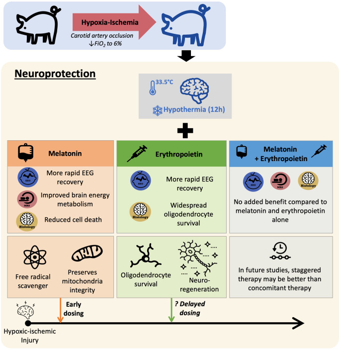
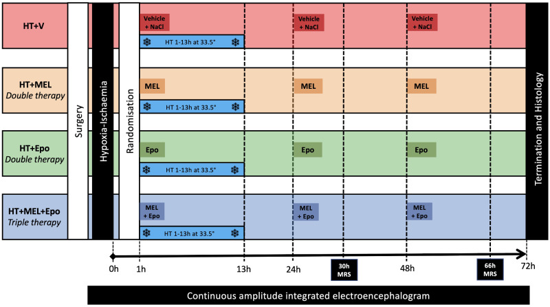
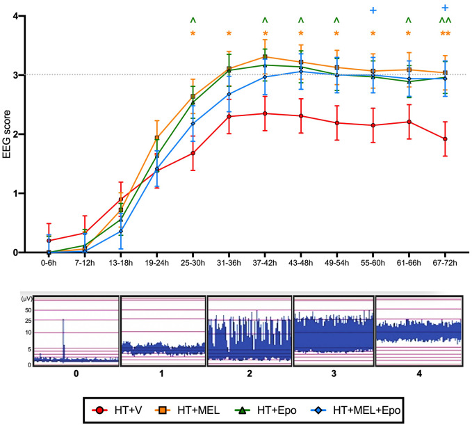
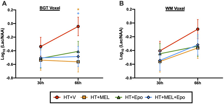
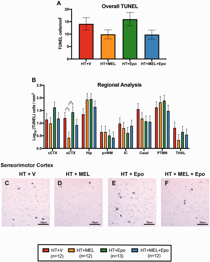
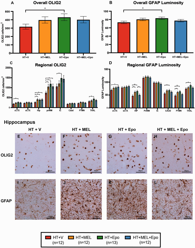
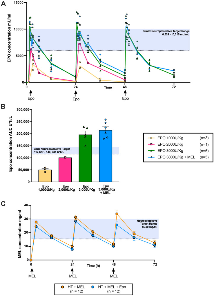
As therapeutic hypothermia is only partially protective for neonatal encephalopathy, safe and effective adjunct therapies are urgently needed. Melatonin and erythropoietin show promise as safe and effective neuroprotective therapies. We hypothesized that melatonin and erythropoietin individually augment 12-h hypothermia (double therapies) and hypothermia + melatonin + erythropoietin (triple therapy) leads to optimal brain protection. Following carotid artery occlusion and hypoxia, 49 male piglets (<48 h old) were randomized to: (i) hypothermia + vehicle (n = 12), (ii) hypothermia + melatonin (20 mg/kg over 2 h) (n = 12), (iii) hypothermia + erythropoietin (3000 U/kg bolus) (n = 13) or (iv) triple therapy (n = 12). Melatonin, erythropoietin or vehicle were given at 1, 24 and 48 h after hypoxia-ischaemia. Hypoxia-ischaemia severity was similar across groups. Therapeutic levels were achieved 3 hours after hypoxia-ischaemia for melatonin (15-30 mg/l) and within 30 min of erythropoietin administration (maximum concentration 10 000 mU/ml). Compared to hypothermia + vehicle, we observed faster amplitude-integrated EEG recovery from 25 to 30 h with hypothermia + melatonin (P = 0.02) and hypothermia + erythropoietin (P = 0.033) and from 55 to 60 h with triple therapy (P = 0.042). Magnetic resonance spectroscopy lactate/N-acetyl aspartate peak ratio was lower at 66 h in hypothermia + melatonin (P = 0.012) and triple therapy (P = 0.032). With hypothermia + melatonin, terminal deoxynucleotidyl transferase-mediated deoxyuridine triphosphate nick-end labelled-positive cells were reduced in sensorimotor cortex (P = 0.017) and oligodendrocyte transcription factor 2 labelled-positive counts increased in hippocampus (P = 0.014) and periventricular white matter (P = 0.039). There was no reduction in terminal deoxynucleotidyl transferase-mediated deoxyuridine triphosphate nick-end labelled-positive cells with hypothermia + erythropoietin, but increased oligodendrocyte transcription factor 2 labelled-positive cells in 5 of 8 brain regions (P < 0.05). Overall, melatonin and erythropoietin were safe and effective adjunct therapies to hypothermia. Hypothermia + melatonin double therapy led to faster amplitude-integrated EEG recovery, amelioration of lactate/N-acetyl aspartate rise and reduction in terminal deoxynucleotidyl transferase-mediated deoxyuridine triphosphate nick-end labelled-positive cells in the sensorimotor cortex. Hypothermia + erythropoietin double therapy was in association with EEG recovery and was most effective in promoting oligodendrocyte survival. Triple therapy provided no added benefit over the double therapies in this 72-h study. Melatonin and erythropoietin influenced cell death and oligodendrocyte survival differently, reflecting distinct neuroprotective mechanisms which may become more visible with longer-term studies. Staggering the administration of therapies with early melatonin and later erythropoietin (after hypothermia) may provide better protection; each therapy has complementary actions which may be time critical during the neurotoxic cascade after hypoxia-ischaemia.

Keywords: erythropoietin; melatonin; neonatal encephalopathy; neuroprotection; therapeutic hypothermia.


Figures

Graphical Abstract
Graphical Abstract
Figure 1
Figure 1
Study protocol. Baseline vital signs were taken prior to induction of anaesthesia and surgical preparation. Following surgery, all piglets underwent cerebral HI by inflation of carotid occluders and reduction of inspired oxygen to 6%. Piglets were resuscitated after HI and observed for 1 h prior to randomization to (i) HT + vehicle (HT+V), (ii) HT+MEL (MEL) double therapy, (iii) HT + Epo double therapy or (iv) HT+MEL+Epo triple therapy. All piglets were cooled to 33.5°C for 12 h from 1 h after HI. MEL 20 mg/kg was infused intravenously over 2 h and Epo 3000 units/kg was given as an intravenous bolus. These were given at 1 h, 24 h and 48 h after HI. The HT+V group received vehicle infusion at an equivalent volume and rate as MEL and a bolus of 0.9% sodium chloride at the same volume as Epo. MRS was acquired out-of-hours at 30 h (18:00) and 66 h (06:00) using the clinical 3 T Philips Achieva magnetic resonance scanner. Cerebral electrical activity was continuously monitored with aEEG. Studies were terminated at 72 h.
Figure 2
Figure 2
aEEG. Hourly aEEG activity was classified according to Hellstrom-Westas et al. (1995) and scores averaged over 6 h intervals. Data presented are the grouped least square means aEEG scores ± standard error of the means (SEM). The least square means was derived from an ANOVA model fitted with fixed factor effects of treatment, time interval and treatment*time interval interaction, plus a random effect subject to take account of repeated measures. Comparison between treatment groups were assessed using 95% confidence intervals for difference in the least square means and P values. Statistical significance when compared to HT+V as shown: *(HT+MEL), ^(HT+Epo) and +(HT+MEL+Epo) where P < 0.05, and **(HT+MEL), ^^(HT+Epo) where P < 0.01.
Figure 3
Figure 3
1H MRS lactate/N-acetyl aspartate (Lac/NAA) peak ratios at 30 h and 66 h after HI. Lac/NAA ratios were obtained for the BGT (A) and WM (B) voxels. An ANOVA model was fitted as previous described. Data presented are the grouped least square means Lac/NAA peak ratio ± standard error of the means (SEM) on the log10 scale. Comparison between treatment groups were assessed using 95% confidence intervals for Log10 (difference in least square means) and P values. Statistical significance shown as *(HT+MEL) and +(HT+MEL+Epo) where P < 0.05 compared to HT+V.
Figure 4
Figure 4
TUNEL immunohistochemistry. Overall (A) and regional (B) TUNEL-positive counts presented as least square means derived from an ANOVA model ± standard error of means (SEM) on the log10 scale. Example micrographs showing TUNEL-positive cells in sCTX with HT+V (C), HT+MEL (D), HT+Epo (E) and HT+MEL+Epo (F). Differences between groups were examined using 95% confidence intervals for the log10 (difference in least square means) and p values. Significant differences shown as *P < 0.05 and **P < 0.01. CAUD = caudate nucleus; cCTX = cingulate gyrus; HIP = hippocampus; IC = internal capsule, PTMN = putamen; PvWM = periventricular white matter; sCTX = sensorimotor cortex; THAL = thalamus.
Figure 5
Figure 5
Immunohistochemistry for oligodendrocytes (OLIG2) and astrocytes (GFAP luminosity). OLIG2 cell counts and GFAP luminosity were fitted to the ANOVA model. The geometric means ± standard error of means (SEM) are shown for the overall (A) and regional (C) OLIG2 cell counts/mm2 after back transformation from the log10 scale. Mean overall (B) and regional (D) GFAP luminosity are also plotted with SEM error bars. Example micrographs from the HIP are shown in (E–L). Differences between groups were examined using 95% confidence intervals for difference in the least square means and significant differences between groups are shown *P < 0.05 and **P < 0.01. CAUD = caudate nucleus; cCTX= cingulate gyrus; HIP = hippocampus; IC = internal capsule; PTMN = putamen; PvWM = periventricular white matter; sCTX = sensorimotor cortex; THAL = thalamus.
Figure 6
Figure 6
Pharmacokinetic (PK) studies of Epo and melatonin in the piglet. Previous pre-clinical studies identified the therapeutic target for Epo neuroprotection as: Cmax 6224–10 015 mU/ml and AUC48 117 677–140 000 U*h/l (Statler et al., 2007). Epo at 1000 u/kg (n = 3), 2000 u/kg (n = 1) and 3000 u/kg [with (n = 5) and without (n = 6) MEL 20 mg/kg)] was administered as an intravenous bolus at 1 h, 24 h and 48 h and Epo levels as maximum concentration (Cmax) (Mean ± SEM) (A) and total AUC levels over 48 h (AUC48) (B) are illustrated. Mean (±standard deviation) melatonin (MEL) plasma concentration (mg/l) for HT+MEL (n = 12) and HT+MEL+Epo (n = 12) treated animals are shown in C. The therapeutic level for MEL is likely between 15 mg/l and 30 mg/l based on our previous studies (Robertson et al., 2013, 2019, 2020).

References

    1. Alonso-Alconada D, Broad KD, Bainbridge A, Chandrasekaran M, Faulkner SD, Kerenyi A, et al. Brain cell death is reduced with cooling by 3.5 degrees C to 5 degrees C but increased with cooling by 8.5 degrees C in a piglet asphyxia model. Stroke 2015; 46: 275–8. - PMC - PubMed
    1. Andrabi SA, Sayeed I, Siemen D, Wolf G, Horn TF.. Direct inhibition of the mitochondrial permeability transition pore: a possible mechanism responsible for anti-apoptotic effects of melatonin. FASEB J 2004; 18: 869–71. - PubMed
    1. Aridas JDS, Yawno T, Sutherland AE, Nitsos I, Ditchfield M, Wong FY, et al. Systemic and transdermal melatonin administration prevents neuropathology in response to perinatal asphyxia in newborn lambs. J Pineal Res 2018; 64: e12479. - PMC - PubMed
    1. Azzopardi D, Chew AT, Deierl A, Huertas A, Robertson NJ, Tusor N, et al. Prospective qualification of early cerebral biomarkers in a randomised trial of treatment with xenon combined with moderate hypothermia after birth asphyxia. EBioMedicine 2019; 47: 484–91. - PMC - PubMed
    1. Azzopardi D, Robertson NJ, Bainbridge A, Cady E, Charles-Edwards G, Deierl A, et al. Moderate hypothermia within 6 h of birth plus inhaled xenon versus moderate hypothermia alone after birth asphyxia (TOBY-Xe): a proof-of-concept, open-label, randomised controlled trial. Lancet Neurol 2016; 15: 145–53. - PMC - PubMed

LinkOut - more resources