Review
. 2021 Jan;478(1):137-150.
doi: 10.1007/s00428-021-03053-1. Epub 2021 Feb 19.

The pulmonary pathology of COVID-19

Affiliations
Review

The pulmonary pathology of COVID-19

Hans Bösmüller et al. Virchows Arch. 2021 Jan.

Abstract

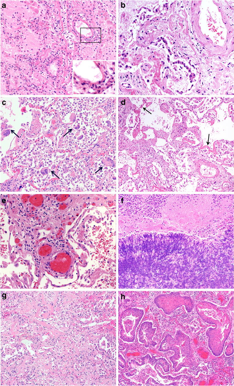
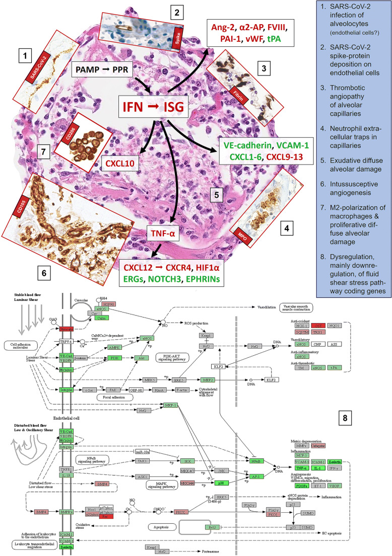
The lung is the main affected organ in severe coronavirus disease 2019 (COVID-19) caused by the novel coronavirus SARS-CoV-2, and lung damage is the leading cause of death in the vast majority of patients. Mainly based on results obtained by autopsies, the seminal features of fatal COVID-19 have been described by many groups worldwide. Early changes encompass edema, epithelial damage, and capillaritis/endothelialitis, frequently combined with microthrombosis. Subsequently, patients with manifest respiratory insufficiency exhibit exudative diffuse alveolar damage (DAD) with hyaline membrane formation and pneumocyte type 2 hyperplasia, variably complicated by superinfection, which may progress to organizing/fibrotic stage DAD. These features, however, are not specific for COVID-19 and can be found in other disorders including viral infections. Clinically, the early disease stage of severe COVID-19 is characterized by high viral load, lymphopenia, massive secretion of pro-inflammatory cytokines and hypercoagulability, documented by elevated D-dimers and an increased frequency of thrombotic and thromboembolic events, whereas virus loads and cytokine levels tend to decrease in late disease stages, when tissue repair including angiogenesis prevails. The present review describes the spectrum of lung pathology based on the current literature and the authors' personal experience derived from clinical autopsies, and tries to summarize our current understanding and open questions of the pathophysiology of severe pulmonary COVID-19.

Keywords: Autopsy; COVID-19; Diffuse alveolar damage; Pulmonary disease; SARS-CoV-2.


Conflict of interest statement

The authors declare no conflict of interest.

Figures

Fig. 1
Fig. 1
Pulmonary pathology of fatal COVID-19. a Incipient pulmonary damage in early-stage disease with intraalveolar edema, endothelial necrosis, microthrombosis, and endothelialitis. Insert with progressive inflammatory vascular injury with nuclear debris and granulocytes. (HE ×100) b Pneumocyte type 2 hyperplasia in exudative phase of DAD. (HE ×100) c Multiple intraalveolar giant cells in exudative phase DAD. (HE ×100) d Hyaline membranes and solid fibrin deposits in exudative phase DAD (HE ×100) e Thrombosis in small- to medium-sized vessels in early-stage lung disease (HE ×100) f Invasive aspergillosis as rare type of superinfection in late-stage DAD. (HE ×100) g Interstitial and intramural fibrosis and intraalveolar plugs of loose connective tissue in organizing phase of DAD. (HE ×100) h Massive bronchopulmonary squamous metaplasia in late stage organizing DAD. (HE ×100)
Fig. 2
Fig. 2
Scheme of the putative pathophysiological mechanisms of SARS-CoV-2 induced acute lung injury, mainly diffuse alveolar damage (background microphotograph (5); H&E, ×200), and microangiopathy and immunopathology in lethal (severe) COVID-19. Inserts, except for (1), are visualized by immunoperoxidase, microphotographed at ×400, and represent (1) SARS-CoV-2 in situ hybridization with the 845701 RNAscope probe - V-nCoV2019-S-sense and visualized with the RNAscope 2.5. LS detection kit (brown) from Advanced Cell Diagnostics (Hayward, CA, USA), yielding linear positivity of an alveolar septum; (2) immunohistochemical staining for SARS-CoV-2 Spike protein with the clone 007 from Sino Biological (Wayne, PA, USA), showing protein deposition on the inner side of an alveolar capillary; (3) immunohistochemical staining for fibrin with a polyclonal antibody A0080 from Dako (Glostrup, Denmark), revealing fibrin microthrombi casting the alveolar capillary network; (4) immunohistochemical staining for myeloperoxidase with a polyclonal antibody 760-2659 from Roche/Ventana (Rotkreuz, Switzerland), with neutrophilic granulocytes stuck into an alveolar capillary and displaying microscopic figures suggestive of neutrophilic extracellular traps (NET); (6) immunohistochemical staining for CD105 with the clone EPR10145-10 from Abcam (Cambridge, UK), showing a tight network of newly formed vessels in an alveolar septum; (7) immunohistochemical staining for CD206 with the clone E2L9N from Cell Signaling (Danvers, MA, USA), with significant amounts of intraalveolar M2 macrophages. (8) Kyoto Encyclopedia of Genes and Genomes (KEGG) diagram of disturbed fluid share stress pathways in COVID-19; respective gene expression profiles have been obtained on 25 lethal COVID-19 cases and compared to lungs of 5 patients suffering from arterial hypertension and 5 histopathologically unremarkable autopsy lungs that served as controls, utilizing the HTG EdgeSeq Oncology Biomarker Panel (HTG Molecular Diagnostics, Tucson, AZ, USA). The scheme should be looked at clockwise from 11 to 7. In general, upregulated genes/proteins are outlined in red, downregulated in green, indifferent ones in black

References

    1. Wu F, Zhao S, Yu B, Chen YM, Wang W, Song ZG, Hu Y, Tao ZW, Tian JH, Pei YY, Yuan ML, Zhang YL, Dai FH, Liu Y, Wang QM, Zheng JJ, Xu L, Holmes EC, Zhang YZ. A new coronavirus associated with human respiratory disease in China. Nature. 2020;579:265–269. doi: 10.1038/s41586-020-2008-3. - DOI - PMC - PubMed
    1. Maiese A, Manetti AC, La Russa R, Di Paolo M, Turillazzi E, Frati P, Fineschi V (2020) Autopsy findings in COVID-19-related deaths: a literature review. Forensic Sci Med Pathol. 10.1007/s12024-020-00310-8 - PMC - PubMed
    1. Omori R, Matsuyama R, Nakata Y. The age distribution of mortality from novel coronavirus disease (COVID-19) suggests no large difference of susceptibility by age. Sci Rep. 2020;10:16642. doi: 10.1038/s41598-020-73777-8. - DOI - PMC - PubMed
    1. Magadum A, Kishore R (2020) Cardiovascular manifestations of COVID-19 infection. Cells 9. 10.3390/cells9112508 - PMC - PubMed
    1. Yang J, Tian C, Chen Y, Zhu C, Chi H, Li J (2020) Obesity aggravates COVID-19: an updated systematic review and meta-analysis. J Med Virol. 10.1002/jmv.26677 - PMC - PubMed