Review
. 2021 Apr;128(4):395-404.
doi: 10.1007/s00702-021-02314-2. Epub 2021 Feb 19.

Dystonia updates: definition, nomenclature, clinical classification, and etiology

Affiliations
Review

Dystonia updates: definition, nomenclature, clinical classification, and etiology

Karen Grütz et al. J Neural Transm (Vienna). 2021 Apr.

Abstract

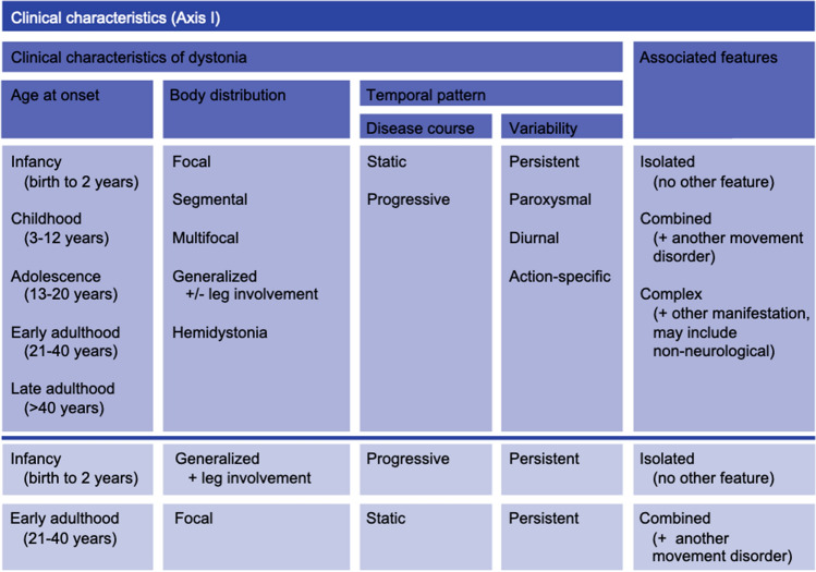
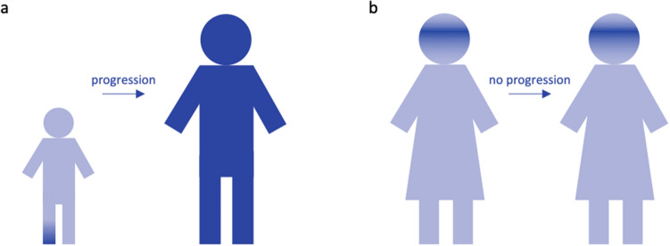
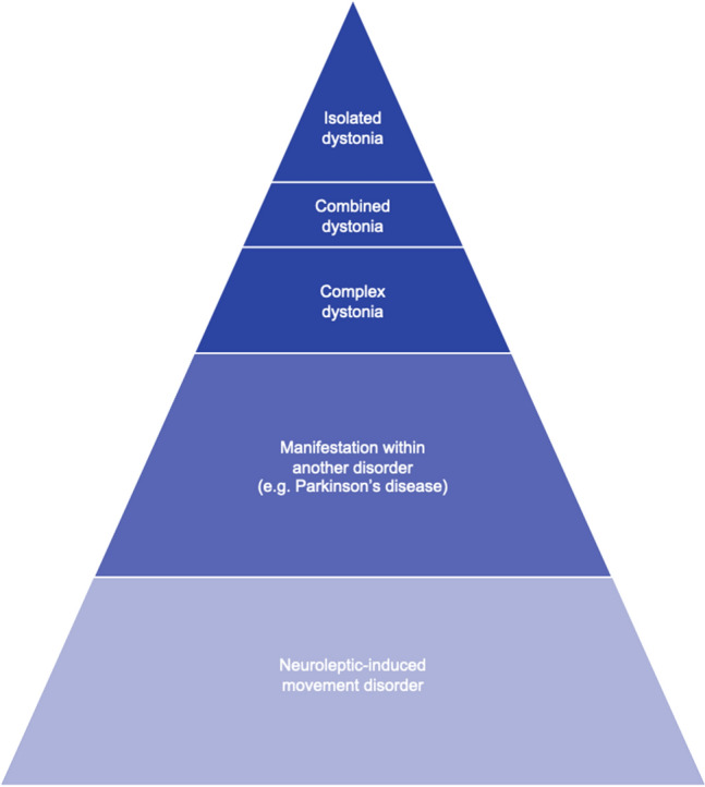
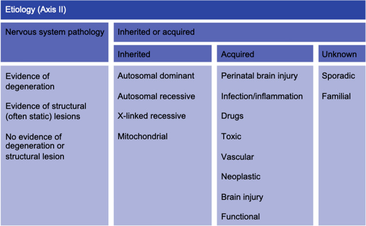
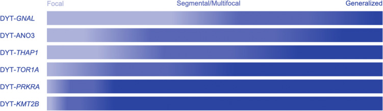
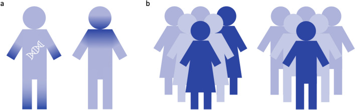
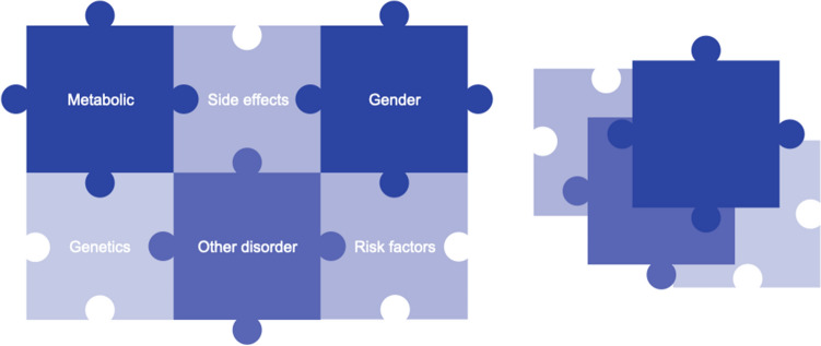
A plethora of heterogeneous movement disorders is grouped under the umbrella term dystonia. The clinical presentation ranges from isolated dystonia to multi-systemic disorders where dystonia is only a co-occurring sign. In the past, definitions, nomenclature, and classifications have been repeatedly refined, adapted, and extended to reflect novel findings and increasing knowledge about the clinical, etiologic, and scientific background of dystonia. Currently, dystonia is suggested to be classified according to two axes. The first axis offers precise categories for the clinical presentation grouped into age at onset, body distribution, temporal pattern and associated features. The second, etiologic, axis discriminates pathological findings, as well as inheritance patterns, mode of acquisition, or unknown causality. Furthermore, the recent recommendations regarding terminology and nomenclature of inherited forms of dystonia and related syndromes are illustrated in this article. Harmonized, specific, and internationally widely used classifications provide the basis for future systematic dystonia research, as well as for more personalized patient counseling and treatment approaches.

Keywords: Clinical classification; Disease etiology; Dystonia; Nomenclature.


Conflict of interest statement

CK serves as a medical advisor to Centogene for genetic testing reports in the fields of movement disorders and dementia, excluding Parkinson’s disease.

Figures

Fig. 1
Fig. 1
Clinical characteristics (Axis I) (adapted from Albanese et al. 2013). In the upper part of the scheme, the four descriptors age at onset, body distribution, temporal pattern, and associated features are depicted with additional categories (temporal pattern) and all recommended subcategories. In the lower part, below the dark blue line, two sets of clinical descriptors are given as examples that are frequently seen in the clinic, although all other combinations of descriptions are possible, as well
Fig. 2
Fig. 2
Disease progression. Dark blue indicates body regions affected with dystonia. Children presenting with focal dystonia have a higher tendency to progress to generalized dystonia (a), while individuals with a late onset, in this case a woman with blepharospasm, will commonly remain stable in their dystonic presentation (b)
Fig.3
Fig.3
Schematic overview on the occurrence of dystonia as a clinical sign according to frequency regardless of the underlying primary condition
Fig. 4
Fig. 4
Etiology (Axis II) (adapted from Albanese et al. 2013). The etiological axis is subdivided into the contribution of the nervous system and the presence of genetic or acquired origin leading to development of dystonia. As an example, an etiological description of a dystonia case can be ‘evidence of degeneration’ and an ‘X-linked recessive inheritance pattern’, as it the case for X-linked dystonia parkinsonism (DYT/PARK-TAF1)
Fig. 5
Fig. 5
Body distribution with respect to genetic background. Different shades of blue indicate the different clinical presentations with darker tones relating to more severe presentations. The fractions are in accordance with the numbers reported by Lange et al. (2021)
Fig. 6
Fig. 6
Distribution of signs and gender differences. a Affected body regions are shown in dark blue. Dystonia of the extremities is more likely to have a genetic origin. b Dark blue represents affected individuals. Women are overall more likely to develop dystonic signs than men
Fig. 7
Fig. 7
Factors contributing to the development and expression of dystonia. Several parts of the puzzle have already been assembled, whereas other pieces of yet unknown entities still need to be added

References

    1. Albanese A, Bhatia K, Bressman SB, et al. Phenomenology and classification of dystonia: a consensus update. Mov Disord. 2013;28:863–873. doi: 10.1002/mds.25475. - DOI - PMC - PubMed
    1. Berman BD, Groth CL, Sillau SH, et al. Risk of spread in adult-onset isolated focal dystonia: a prospective international cohort study. J Neurol Neurosurg Psychiatry. 2020;91:314–320. doi: 10.1136/jnnp-2019-321794. - DOI - PMC - PubMed
    1. Bressman SB, Sabatti C, Raymond D, et al. The DYT1 phenotype and guidelines for diagnostic testing. Neurology. 2000;54:1746–1752. doi: 10.1212/wnl.54.9.1746. - DOI - PubMed
    1. Bull PC, Thomas GR, Rommens JM, et al. The Wilson disease gene is a putative copper transporting P-type ATPase similar to the menkes gene. Nat Genet. 1993;5:327–337. doi: 10.1038/ng1293-327. - DOI - PubMed
    1. Cascalho A, Foroozandeh J, Hennebel L, et al. Excess Lipin enzyme activity contributes to TOR1A recessive disease and DYT-TOR1A dystonia. Brain J Neurol. 2020;143:1746–1765. doi: 10.1093/brain/awaa139. - DOI - PubMed

Publication types