. 2021 Mar 1;218(3):e20201006.
doi: 10.1084/jem.20201006.

LACC1 deficiency links juvenile arthritis with autophagy and metabolism in macrophages

Affiliations

LACC1 deficiency links juvenile arthritis with autophagy and metabolism in macrophages

Ommar Omarjee et al. J Exp Med. .

Abstract

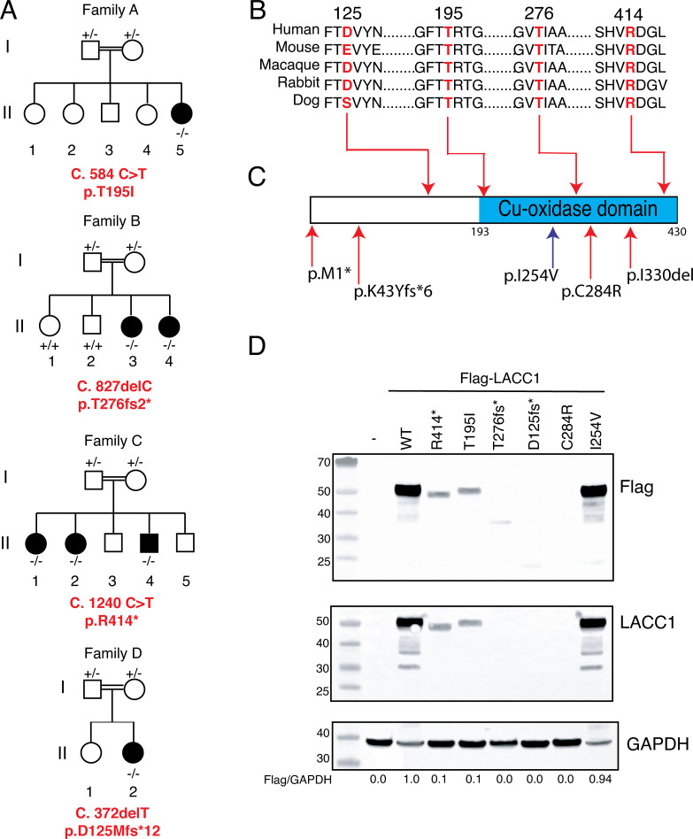
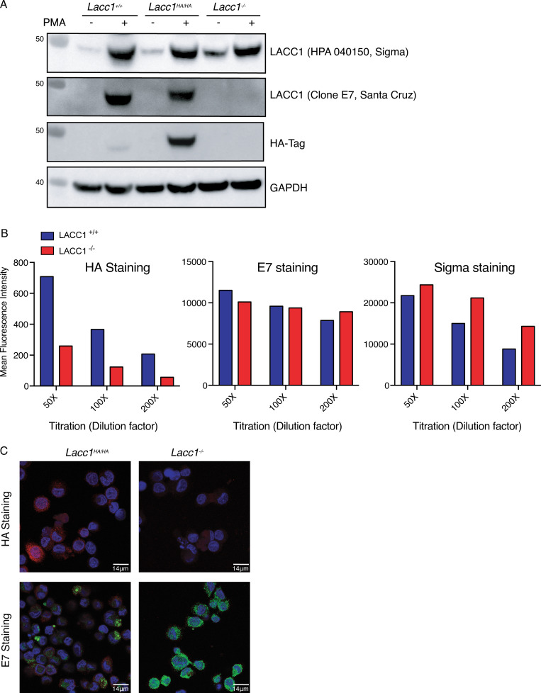
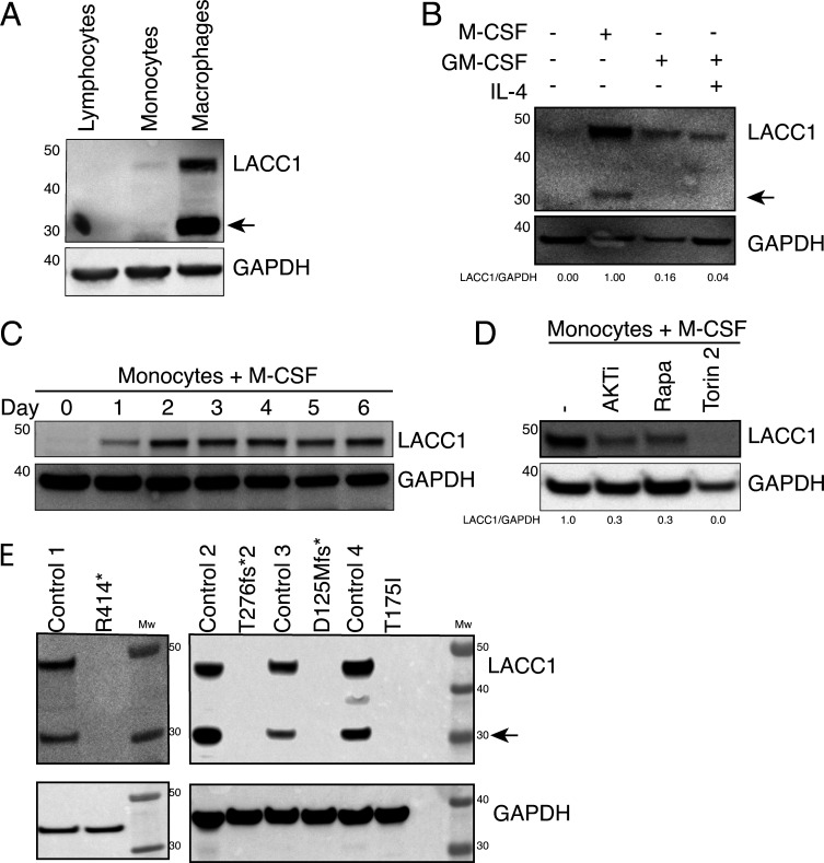
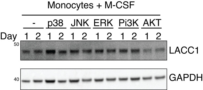
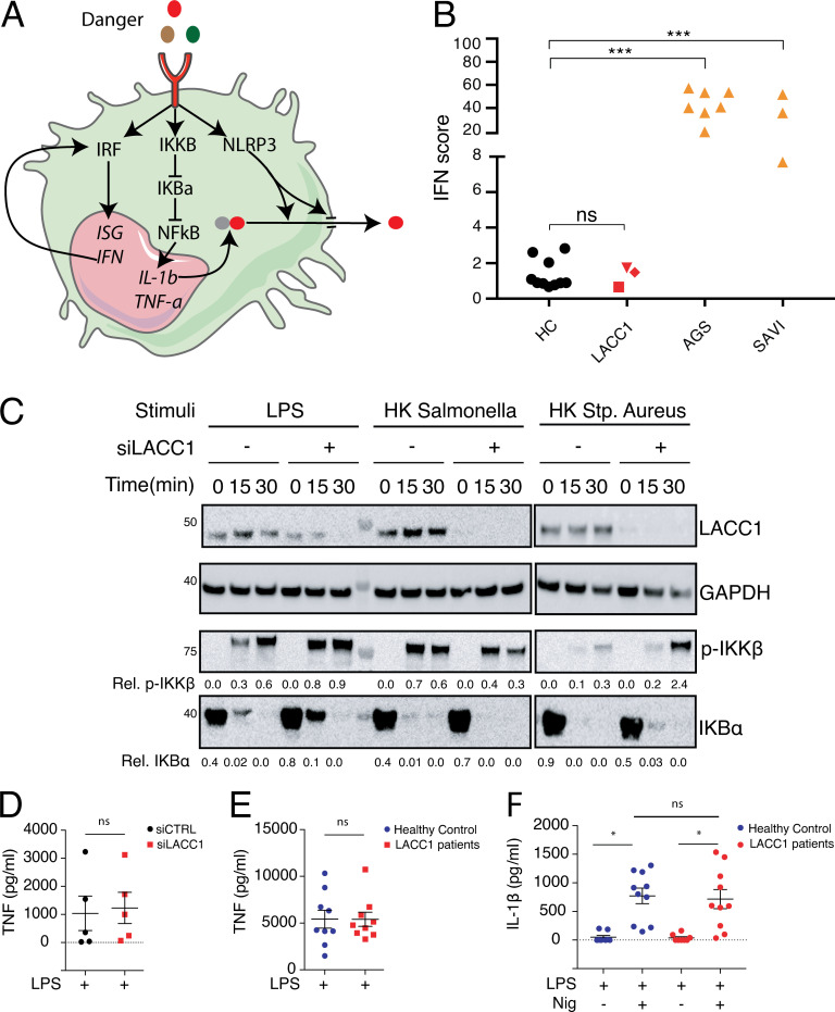
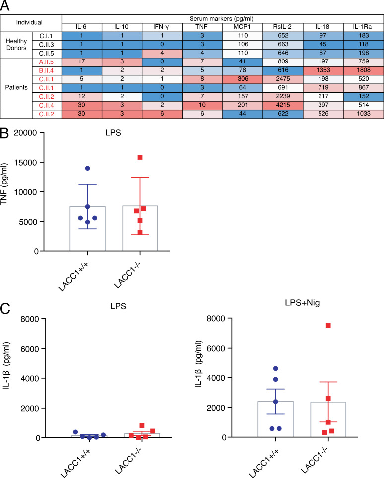
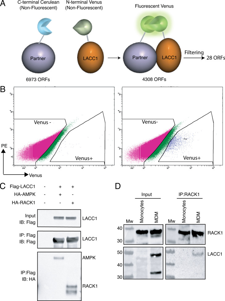
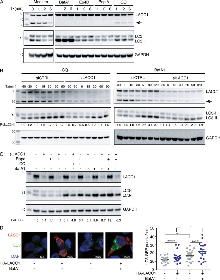
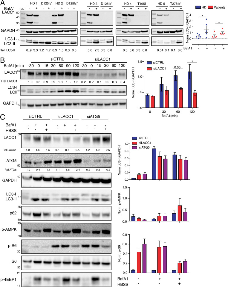
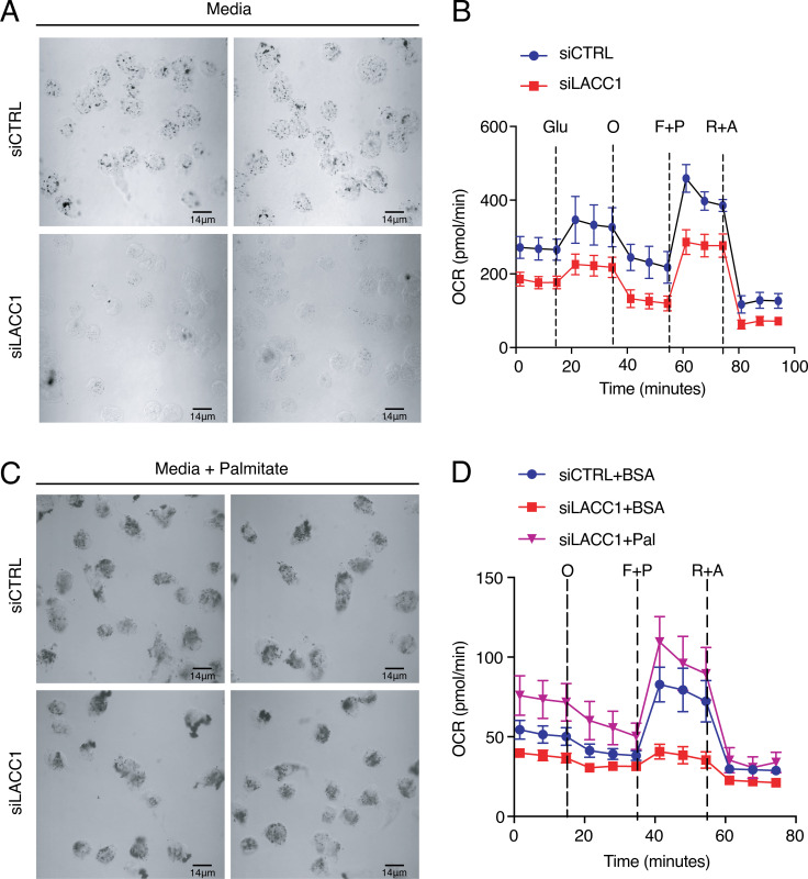
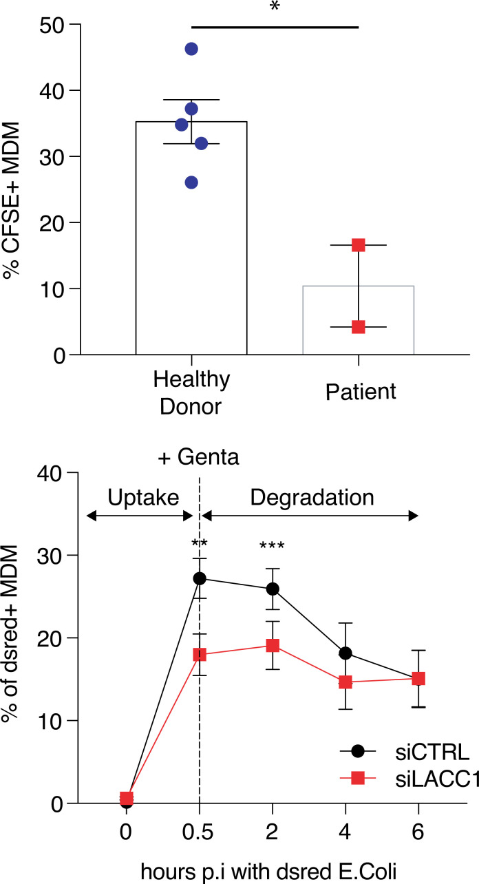
Juvenile idiopathic arthritis is the most common chronic rheumatic disease in children, and its etiology remains poorly understood. Here, we explored four families with early-onset arthritis carrying homozygous loss-of-expression mutations in LACC1. To understand the link between LACC1 and inflammation, we performed a functional study of LACC1 in human immune cells. We showed that LACC1 was primarily expressed in macrophages upon mTOR signaling. We found that LACC1 deficiency had no obvious impact on inflammasome activation, type I interferon response, or NF-κB regulation. Using bimolecular fluorescence complementation and biochemical assays, we showed that autophagy-inducing proteins, RACK1 and AMPK, interacted with LACC1. Autophagy blockade in macrophages was associated with LACC1 cleavage and degradation. Moreover, LACC1 deficiency reduced autophagy flux in primary macrophages. This was associated with a defect in the accumulation of lipid droplets and mitochondrial respiration, suggesting that LACC1-dependent autophagy fuels macrophage bioenergetics metabolism. Altogether, LACC1 deficiency defines a novel form of genetically inherited juvenile arthritis associated with impaired autophagy in macrophages.


Conflict of interest statement

Disclosures: S. Georgin-Lavialle reported personal fees from SOBI, non-financial support from Novartis, and personal fees from BMS outside the submitted work. F. Bleicher reported a patent to FR1655539 pending. J. Reboulet reported a patent to FR1655539 issued. S. Merabet reported a patent to FR1655539 pending. T. Henry reported personal fees from SOBI and grants from SOBI outside the submitted work. No other disclosures were reported.

Figures

Figure 1.
Figure 1.
Whole or targeted exome sequencing identifies loss-of-function mutations in LACC1 from JIA families. (A) Pedigree of four JIA families with identified LACC1 mutations. (B) Protein alignment at mutated amino acids sites across species. (C) Representation of novel JIA mutations (upper red arrows), previously reported JIA mutations (lower red arrow), and Crohn’s disease–associated variant (lower blue arrow) on the structure of LACC1 with a predicted copper-oxidase domain. (D) Immunoblots of 293T cells transfected with plasmids encoding for flagged forms of LACC1 WT or variants (representative of two experiments).
Figure S1.
Figure S1.
Endogenous LACC1 tagging reveals unspecific staining by previously used commercial antibodies. (A) Immunoblots of U937 either WT, HA-tagged, or deficient for LACC1 using anti-LACC1 (E7 clone; Santa Cruz), anti-LACC1 (HPA040150; Sigma-Aldrich), or anti-HA (901524; Biolegend) antibody. (B) Intracellular FACS staining of U937 HA-LACC1 or LACC1−/− with indicated antibodies against LACC1. (C) Confocal analysis of U937 HA-LACC1 or LACC1−/− with indicated antibodies against LACC1. Nucleus was stained with DAPI (blue).
Figure 2.
Figure 2.
LACC1 is strongly expressed in an mTOR-dependent manner during M-CSF–induced human monocyte–macrophage differentiation. (A) Immunoblots of various immune cells isolated from buffy coat (lymphocyte and monocytes) or differentiated in vitro (M-CSF macrophages; representative of three experiments). Arrowhead indicates short fragment of LACC1. (B) Immunoblot of monocytes stimulated with the indicated cytokines for 6 d (representative of two experiments). Arrowhead indicates short fragment of LACC1. (C) Immunoblots of monocytes treated with M-CSF at different time points (representative of three experiments). (D) Immunoblots of monocytes treated with M-CSF and inhibitors (AKTi, Rapamycin, and Torin2) over 24 h (representative of two experiments). (E) Immunoblots of macrophages derived from either four healthy donors or four JIA patients bearing indicated mutations in LACC1. Mw, molecular weight. For B and D, a quantification of LACC1 level is shown below the blot.
Figure S2.
Figure S2.
Role of major signaling pathways on LACC1 expression in macrophages. Immunoblots of monocytes treated with or without M-CSF and different inhibitors of p38, JNK, ERK, PI3K, and AKT during the indicated period (representative of two experiments).
Figure 3.
Figure 3.
LACC1 deficiency does not seem to be connected to known monogenic autoinflammatory pathways. (A) Illustration of IFN, NF-κB, and inflammasome activation pathways. IRF, IFN regulatory factor. (B) IFN score determined from Nanostring measurement of ISGs in whole blood of 10 controls and patients C.II.1, C.II.2, and C.II.4. Samples from Aicardi–Goutières syndrome (AGS) and STING-associated vasculopathy of infancy patients (SAVI) were used as controls. ***, P < 0.0005 (unpaired t test). (C) Immunoblots (IB) of indicated siRNA-transfected human monocyte–derived macrophages stimulated with LPS (n = 3), heat-killed (HK) Salmonella, or heat-killed S. (Stp.) aureus (n = 2) for 15 and 30 min. (D) TNF secretion of human monocyte–derived macrophages stimulated with LPS for 3 h (n = 4). (E) TNF secretion of monocyte-derived macrophages obtained from healthy donors and LACC1 patients stimulated with LPS for 3 h (n = 9). (F) IL-1β secretion of monocyte-derived macrophages obtained from healthy donors and LACC1 patients stimulated with LPS ± nigericine (Nig; n = 10). *, P < 0.05 (paired t test).
Figure S3.
Figure S3.
Inflammatory cytokine production in a LACC1-deficient environment. (A) Heatmap representation of serum cytokine levels of three healthy family members (black) and seven LACC1 patients (red). (B) U937 were differentiated for 48 h with PMA and stimulated 3 h with LPS (n = 5). Supernatants were assayed for TNF levels. (C) U937 were differentiated for 48 h with PMA and stimulated 3 h with LPS ± Nigericin (Nig; n = 5). Supernatant were assayed for IL-1β levels.
Figure 4.
Figure 4.
A large-scale proteomic screen identifies AMPK and RACK1 as LACC1 partners. (A) Schematic representation of proteomic screen design consisting of the cotransfection of LACC1 and ORF library tagged with either fragments of Venus protein. (B) GFP-positive cell sorting of 293T cells expressing BiFC ORFs transfected with (right) or without (left) N-Venus-LACC1. (C) Immunoblots of Flag-immunoprecipitated cell lysates of 293T cells cotransfected with tagged LACC1, AMPK, or RACK1 expression vectors (representative of three experiments). (D) Immunoblots of endogenous RACK1-immunoprecipitated cell lysates of human monocytes and macrophages. Input, immunoprecipitated (IP) fractions were immunoblotted against LACC1 (representative of two experiments). MDM, monocyte-derived macrophage; Mw, molecular weight.
Figure S4.
Figure S4.
Autophagy-lysosome inhibitors control LACC1 cleavage and confirms LC3-II accumulation defect in the absence of LACC1. (A) Immunoblots of M-CSF macrophages treated with or without lysosome-autophagy inhibitors such as Bafilomycin A1 (BafA1), cathepsin inhibitors E64D and pepstatin A (Pep A) or chloroquine (CQ) at different time points (Tx). (B) Immunoblots of siLACC1 macrophages treated with either chloroquine (n = 2) or BafA1 (n = 3). (C) Immunoblots of siLACC1 macrophages treated with a combination of rapamycin (Rapa), chloroquine, or BafA1 for 2 h (n = 2). (D) Confocal microscopy of HeLa cells stably expressing GFP-LC3 (green) transfected with Flag-LACC1 (red). Nucleus was stained with DAPI (blue; representative of three experiments). Right: Quantification of the number of LC3-GFP dots per cell in the different conditions. *, P < 0.05; **, P < 0.005.
Figure 5.
Figure 5.
LACC1 regulates autophagy and associated signaling. (A) Immunoblot of five healthy donors (HD) versus five patients (three different samplings of D125fs* [triangle], T196I [circle], and T276fs* [star]) macrophages treated with BafA1 for 2 h. Right: LC3-II levels normalized to GAPDH. (*, P < 0.05 [paired t test]).(B) Immunoblot of control versus siLACC1 macrophages treated with BafA1 at different time points (representative of three experiments). Right: Accumulation of LC3-II levels normalized to GAPDH at selected time points based on the immunoblots. *, P < 0.05 (paired t test). (C) Immunoblot of control versus LACC1-deficient macrophages treated with BafA1 in RPMI or HBSS starvation medium for 2 h (representative of two experiments). LACC1 and ATG5 levels have been normalized with respect to GAPDH and unstimulted siCTRL condition. The right panels indicate normalized LC3-II (upper), phospho-APMK (middle), and phospho-S6 (lower) to GAPDH. CTRL, control; Norm, normal.
Figure 6.
Figure 6.
LACC1 promotes lipid droplet and mitochondrial respiration. (A) Red oil staining of macrophages transfected with control or LACC1 siRNA in basal RPMI medium (representative of three experiments). (B) OCR measurement of macrophages transfected with control or LACC1 siRNA with successive glucose (Glu), oligomycin (O), FCCP (F+P), and rotenone and antimycin A (R+A) treatments (representative of three experiments). (C) Red oil staining of macrophages transfected with control or LACC1 siRNA in palmitate supplemented medium for 16 h (representative of two experiments). (D) OCR measurement of transfected with control or LACC1 siRNA treated with palmitate followed by successive addition of glucose, oligomycin, FCCP, and rotenone and antimycin A (representative of two experiments). Pal, palmitate.
Figure S5.
Figure S5.
LACC1-deficient macrophages have impaired capacity to clear apoptotic bodies and bacteria. (A) FACS analysis of five control or two LACC1 patients’ macrophages preincubated for 4 h with CFSE-stained apoptotic bodies. (B) Control or siLACC1 macrophages were preincubated for 30 min with live DsRed E. coli before removal and addition of sterile medium containing antibiotic. Macrophages were analyzed at different time points post infection (p.i.) for bacterial uptake (n = 4; *, P < 0.05; paired t test). MDM, monocyte-derived macrophage.

Comment in

References

    1. Aksentijevich, I., and Zhou Q.. 2017. NF-κB Pathway in Autoinflammatory Diseases: Dysregulation of Protein Modifications by Ubiquitin Defines a New Category of Autoinflammatory Diseases. Front. Immunol. 8:399. 10.3389/fimmu.2017.00399 - DOI - PMC - PubMed
    1. Assadi, G., Saleh R., Hadizadeh F., Vesterlund L., Bonfiglio F., Halfvarson J., Törkvist L., Eriksson A.S., Harris H.E., Sundberg E., and D’Amato M.. 2016. LACC1 polymorphisms in inflammatory bowel disease and juvenile idiopathic arthritis. Genes Immun. 17:261–264. 10.1038/gene.2016.17 - DOI - PubMed
    1. Baldassano, R.N., Bradfield J.P., Monos D.S., Kim C.E., Glessner J.T., Casalunovo T., Frackelton E.C., Otieno F.G., Kanterakis S., Shaner J.L., et al. . 2007. Association of the T300A non-synonymous variant of the ATG16L1 gene with susceptibility to paediatric Crohn’s disease. Gut. 56:1171–1173. 10.1136/gut.2007.122747 - DOI - PMC - PubMed
    1. Bonilla, D.L., Bhattacharya A., Sha Y., Xu Y., Xiang Q., Kan A., Jagannath C., Komatsu M., and Eissa N.T.. 2013. Autophagy regulates phagocytosis by modulating the expression of scavenger receptors. Immunity. 39:537–547. 10.1016/j.immuni.2013.08.026 - DOI - PMC - PubMed
    1. Cader, M.Z., Boroviak K., Zhang Q., Assadi G., Kempster S.L., Sewell G.W., Saveljeva S., Ashcroft J.W., Clare S., Mukhopadhyay S., et al. . 2016. C13orf31 (FAMIN) is a central regulator of immunometabolic function. Nat. Immunol. 17:1046–1056. 10.1038/ni.3532 - DOI - PMC - PubMed

Publication types

MeSH terms