Clinical Trial
. 2021 Feb 19;16(2):e0244781.
doi: 10.1371/journal.pone.0244781. eCollection 2021.

Sonographic findings in coronavirus disease-19 associated liver damage

Affiliations
Clinical Trial

Sonographic findings in coronavirus disease-19 associated liver damage

Jakob Spogis et al. PLoS One. .

Abstract

Purpose: This study was conducted to evaluate the role of liver sonography in patients with coronavirus disease 2019 (COVID-19) and elevated liver enzymes.

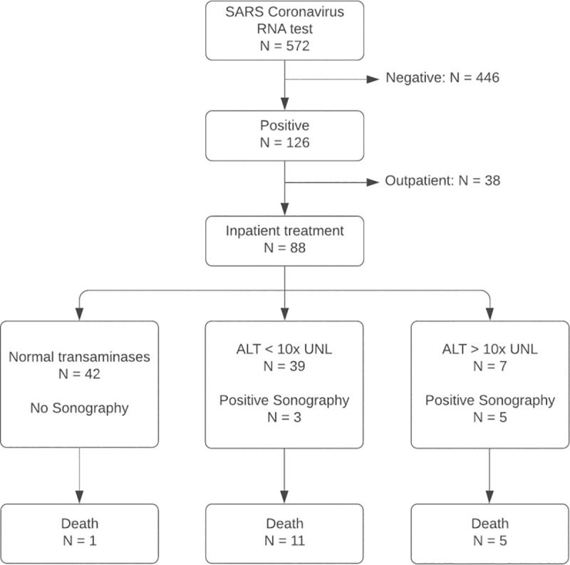
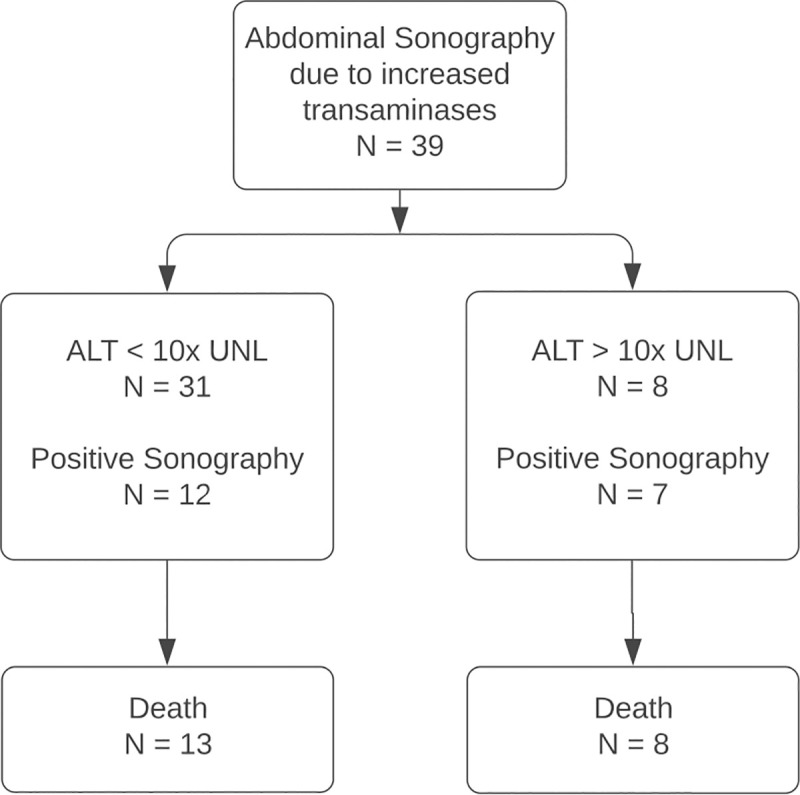
Materials and methods: In this retrospective study, patients tested positive for SARS-CoV-2 in our emergency ward between January 01 and April 24, 2020 and elevated liver enzymes were included (Cohort 1). Additionally, the local radiology information system was screened for sonographies in COVID-19 patients at the intensive care unit in the same period (Cohort 2). Liver sonographies and histologic specimen were reviewed and suspicious findings recorded. Medical records were reviewed for clinical data. Ultrasound findings and clinical data were correlated with severity of liver enzyme elevation.

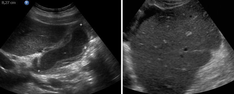
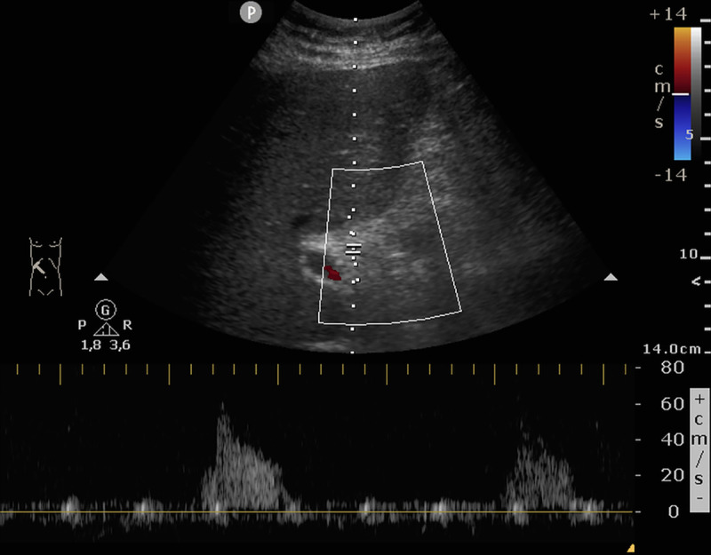
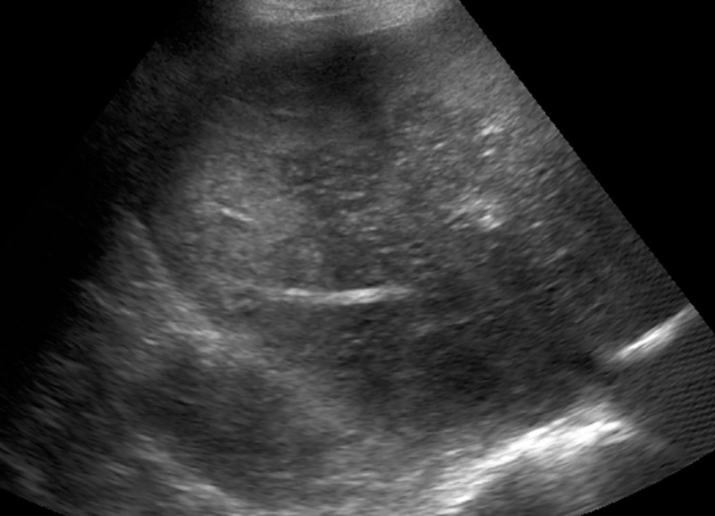
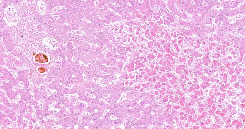
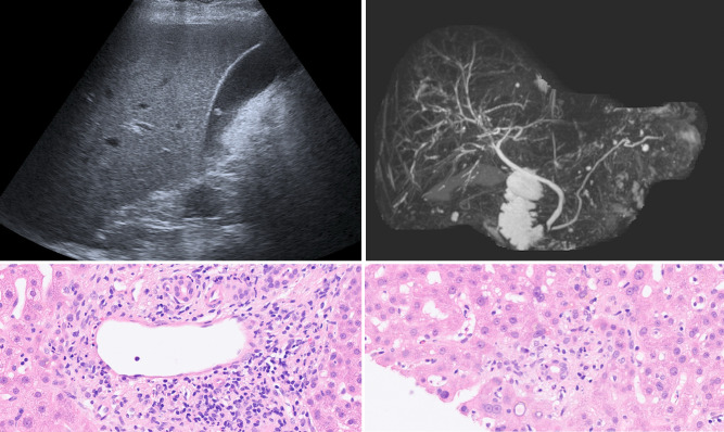
Results: Cohort 1: 126 patients were evaluated, of which 47 (37.3%) had elevated liver enzymes. Severity of liver enzyme elevation was associated with death (p<0.001). 8 patients (6.3%) had suspicious ultrasound findings, including signs of acute hepatitis (n = 5, e.g. thickening of gall bladder wall, hepatomegaly, decreased echogenicity of liver parenchyma) and vascular complications (n = 4). Cohort 2: 39 patients were evaluated, of which 14 are also included in Cohort 1. 19 patients (48.7%) had suspicious ultrasound findings, of which 13 patients had signs of acute hepatitis and 6 had vascular complications. Pathology was performed in 6 patients. Predominant findings were severe cholestasis and macrophage activation.

Conclusion: For most hospitalized COVID-19 patients, elevated liver enzymes cause little concern as they are only mild to moderate. However, in severely ill patients bedside sonography is a powerful tool to reveal different patterns of vascular, cholestatic or inflammatory complications in the liver, which are associated with high mortality. In addition, macrophage activation as histopathologic correlate for a hyperinflammatory syndrome seems to be a frequent complication in COVID-19.


Conflict of interest statement

The authors have declared that no competing interests exist.

Figures

Fig 1
Fig 1. Procedure for research subject selection of cohort 1.
All patients positively tested for SARS Coronavirus RNA were screened for elevated liver enzymes. ALT, alanine aminotransferase; UNL, upper normal limit.
Fig 2
Fig 2. Procedure for research subject selection of cohort 2.
The local radiology information system (RIS) was screened for abdominal sonographies at the intensive care unit for diagnostic workup of elevated liver enzymes. ALT, alanine aminotransferase; UNL, upper normal limit.
Fig 3
Fig 3. Signs of acute hepatitis.
(A) Pronounced thickening of the gallbladder wall, filled with sludge. This represents a typical sonographic finding in our study. (B) Indicated starry sky appearance of the liver with decreased echogenicity of liver parenchyma and pronounced bile ducts.
Fig 4
Fig 4. Vascular complications in the liver.
In this patient with severe deterioration of liver function only residual arterial Doppler signal could be found in the porta hepatis. The patient died the same day.
Fig 5
Fig 5. Vascular complications in the liver.
In two cases, inhomogeneous echogenicity of the liver parenchyma was found. Autopsy confirmed the suspected diagnosis of liver necrosis in this case.
Fig 6
Fig 6. Histologic specimen of the liver.
Progressive centroacinar hypoxic damage (right) and conspicuous cholestasis in adjacent portal field. HE x200.
Fig 7
Fig 7. Patient of group 3 with persistent severe elevation of liver enzymes and GGT.
(A) Whereas sonography was normal, (B) MRCP revealed irregular bile ducts leading to the suspected diagnosis of secondary sclerosing cholangitis (SSC). (C, D): Histologic specimen of the same patient showing signs of vascular- (C) and bile duct-associated (D) inflammation, which is typical for misguided immunoreaction like in rejection reaction. The diagnosis of SSC could not be confirmed.

References

    1. Guan WJ, Ni ZY, Hu Y, Liang WH, Ou CQ, He JX, et al. Clinical Characteristics of Coronavirus Disease 2019 in China. N Engl J Med. 2020;382(18):1708–20. Epub 2020/02/29. 10.1056/NEJMoa2002032 - DOI - PMC - PubMed
    1. Bangash MN, Patel J, Parekh D. COVID-19 and the liver: little cause for concern. Lancet Gastroenterol Hepatol. 2020;5(6):529–30. Epub 2020/03/24. 10.1016/S2468-1253(20)30084-4 . - DOI - PMC - PubMed
    1. Li X, Wang L, Yan S, Yang F, Xiang L, Zhu J, et al. Clinical characteristics of 25 death cases with COVID-19: A retrospective review of medical records in a single medical center, Wuhan, China. Int J Infect Dis. 2020;94:128–32. Epub 2020/04/07. 10.1016/j.ijid.2020.03.053 - DOI - PMC - PubMed
    1. Xu Z, Shi L, Wang Y, Zhang J, Huang L, Zhang C, et al. Pathological findings of COVID-19 associated with acute respiratory distress syndrome. Lancet Respir Med. 2020;8(4):420–2. Epub 2020/02/23. 10.1016/S2213-2600(20)30076-X - DOI - PMC - PubMed
    1. Sun J, Aghemo A, Forner A, Valenti L. COVID-19 and liver disease. Liver Int. 2020;40(6):1278–81. Epub 2020/04/07. 10.1111/liv.14470 . - DOI - PubMed

Publication types