. 2021 Feb 19;16(2):e0246764.
doi: 10.1371/journal.pone.0246764. eCollection 2021.

Left ventricular myocardial strain and tissue characterization by cardiac magnetic resonance imaging in immune checkpoint inhibitor associated cardiotoxicity

Affiliations

Left ventricular myocardial strain and tissue characterization by cardiac magnetic resonance imaging in immune checkpoint inhibitor associated cardiotoxicity

Angela Y Higgins et al. PLoS One. .

Abstract

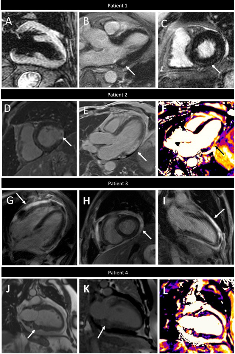
Background: Immune checkpoint inhibitors (ICIs) are highly effective in treating cancer; however, cardiotoxicity can occur, including myocarditis. Cardiac magnetic resonance (CMR) imaging is useful for evaluation of myocarditis, although it has not been well studied in ICI cardiotoxicity.

Methods: We identified patients referred for CMR evaluation of ICI cardiotoxicity from September 2015 through September 2019. We assessed structural and functional parameters, feature tracking (FT) left ventricular and atrial strain, T2- weighted ratios and quantitative late gadolinium enhancement (LGE). We also applied the Updated Lake Louise Criteria for diagnosis of myocarditis.

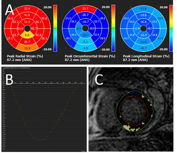
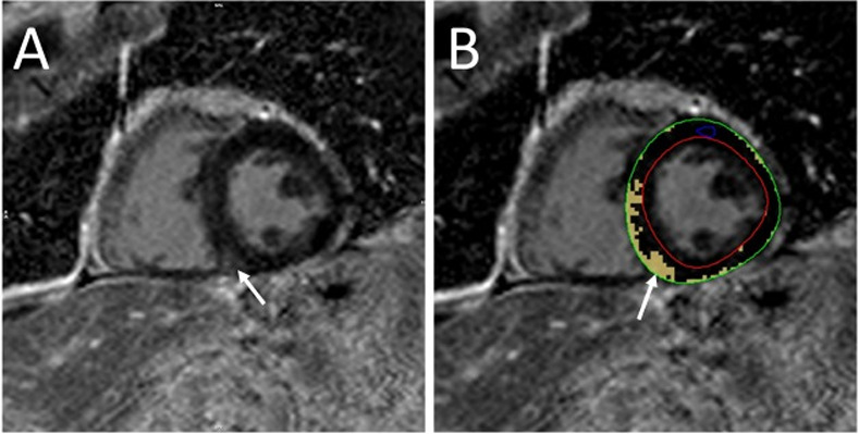
Results: Of the 20 patients referred, the median left ventricular ejection fraction (LVEF) was 52.5% ± 19.1 and 50% had a normal LVEF (≥53%). FT strain analysis revealed an average abnormal global longitudinal strain (GLS) of -9.8%± 4.2%. In patients with a normal LVEF, the average GLS remained depressed at -12.3%± 2.4%. In all patients, GLS demonstrated a significant negative correlation with LVEF (rs = -0.64, p 0.002). Sixteen patients (80%) had presence of LGE (14 non-ischemic pattern and 2 ischemic). Percent LGE did not correlate with any CMR parameters and notably did not correlate with LVEF (rs = -0.29, p = 0.22) or GLS (rs = 0.10, p = 0.67), highlighting the value of tissue characterization beyond functional assessment. Nine patients (45%) met full Updated Lake Louise Criteria and 85% met at least one criterion, suggestive of myocarditis in the correct clinical context. Thirteen patients (65%) were treated for ICI-associated myocarditis and, of these, 54% (n = 7) had recovery of LVEF to normal. There was no correlation between LVEF (p = 0.47), GLS (0.89), or % LGE (0.15) and recovery of LVEF with treatment.

Conclusion: In patients with suspected ICI cardiotoxicity, CMR is an important diagnostic tool, even in the absence of overt left ventricular dysfunction, as abnormalities in left ventricular strain, T2 signal and LGE can identifying disease.


Conflict of interest statement

The authors have declared that no competing interests exist.

Figures

Fig 1
Fig 1. Sample CMR strain analysis.
CMR showed normal LVEF (53%) with abnormal strain: A. peak global radial strain 36.4%, circumferential strain -19.7% and longitudinal strain −13.8%, B. graphical representation of peak global longitudinal strain and C. quantitative LGE of 7%.
Fig 2
Fig 2. Sample quantitative late gadolinium enhancement.
Mid-myocardial LGE of the basal-to-mid inferoseptal wall and inferior right ventricular insertion site (A) and quantitative scar 8.4% (B).
Fig 3
Fig 3. Time from starting Immune Checkpoint Inhibitor (ICI) to clinical presentation.
Fig 4
Fig 4. Distribution of primary symptom on presentation (n = 20).
Fig 5
Fig 5. Varying CMR presentations of Immune Checkpoint Inhibitor (ICI) associated cardiotoxicity.
Patient 1. Myocarditis with elevated T2-weighted signal (T2 ratio 2.2) (A) three chamber (B, white arrow) and short axis (C, white arrow) LGE imaging showing mid-myocardial LGE of basal inferior and inferoseptal walls. Patient 2. Acute myocardial infarction showing transmural LGE of the inferolateral wall with microvascular obstruction (D and E, white arrows) and corresponding increased signal on T2W-mapping (F, black arrow). Patient 3. Myopericarditis showing pericardial thickening and LGE on four chamber (G), short axis (H) and two chamber (I). Patient 4. Metastatic tumors within the basal inferior myocardium on two-chamber cine imaging (J, white arrow) with corresponding LGE (I, arrow) and edema on T2W imaging (J, black arrow).

Similar articles

Cited by

References

    1. Tang J, Yu JX, Hubbard-Lucey VM, Neftelinov ST, Hodge JP, Lin Y. Trial watch: The clinical trial landscape for PD1/PDL1 immune checkpoint inhibitors. Nat Rev Drug Discov. 2018;17(12):854–5. 10.1038/nrd.2018.210 - DOI - PubMed
    1. De Velasco G, Je Y, Bosse D, Awad MM, Ott PA, Moreira RB, et al. Comprehensive Meta-analysis of Key Immune-Related Adverse Events from CTLA-4 and PD-1/PD-L1 Inhibitors in Cancer Patients. Cancer Immunol Res. 2017;5(4):312–8. 10.1158/2326-6066.CIR-16-0237 - DOI - PMC - PubMed
    1. Mahmood SS, Fradley MG, Cohen JV, Nohria A, Reynolds KL, Heinzerling LM, et al. Myocarditis in Patients Treated With Immune Checkpoint Inhibitors. J Am Coll Cardiol. 2018;71(16):1755–64. 10.1016/j.jacc.2018.02.037 - DOI - PMC - PubMed
    1. Zhou YW, Zhu YJ, Wang MN, Xie Y, Chen CY, Zhang T, et al. Immune Checkpoint Inhibitor-Associated Cardiotoxicity: Current Understanding on Its Mechanism, Diagnosis and Management. Front Pharmacol. 2019;10:1350 10.3389/fphar.2019.01350 - DOI - PMC - PubMed
    1. Wang Y, Zhou S, Yang F, Qi X, Wang X, Guan X, et al. Treatment-Related Adverse Events of PD-1 and PD-L1 Inhibitors in Clinical Trials: A Systematic Review and Meta-analysis. JAMA Oncol. 2019;5(7):1008–19. 10.1001/jamaoncol.2019.0393 - DOI - PMC - PubMed

Publication types

MeSH terms

LinkOut - more resources