. 2021 Feb 18;81(4):739-755.e7.
doi: 10.1016/j.molcel.2021.01.024.

cGAS phase separation inhibits TREX1-mediated DNA degradation and enhances cytosolic DNA sensing

Affiliations

cGAS phase separation inhibits TREX1-mediated DNA degradation and enhances cytosolic DNA sensing

Wen Zhou et al. Mol Cell. .

Abstract

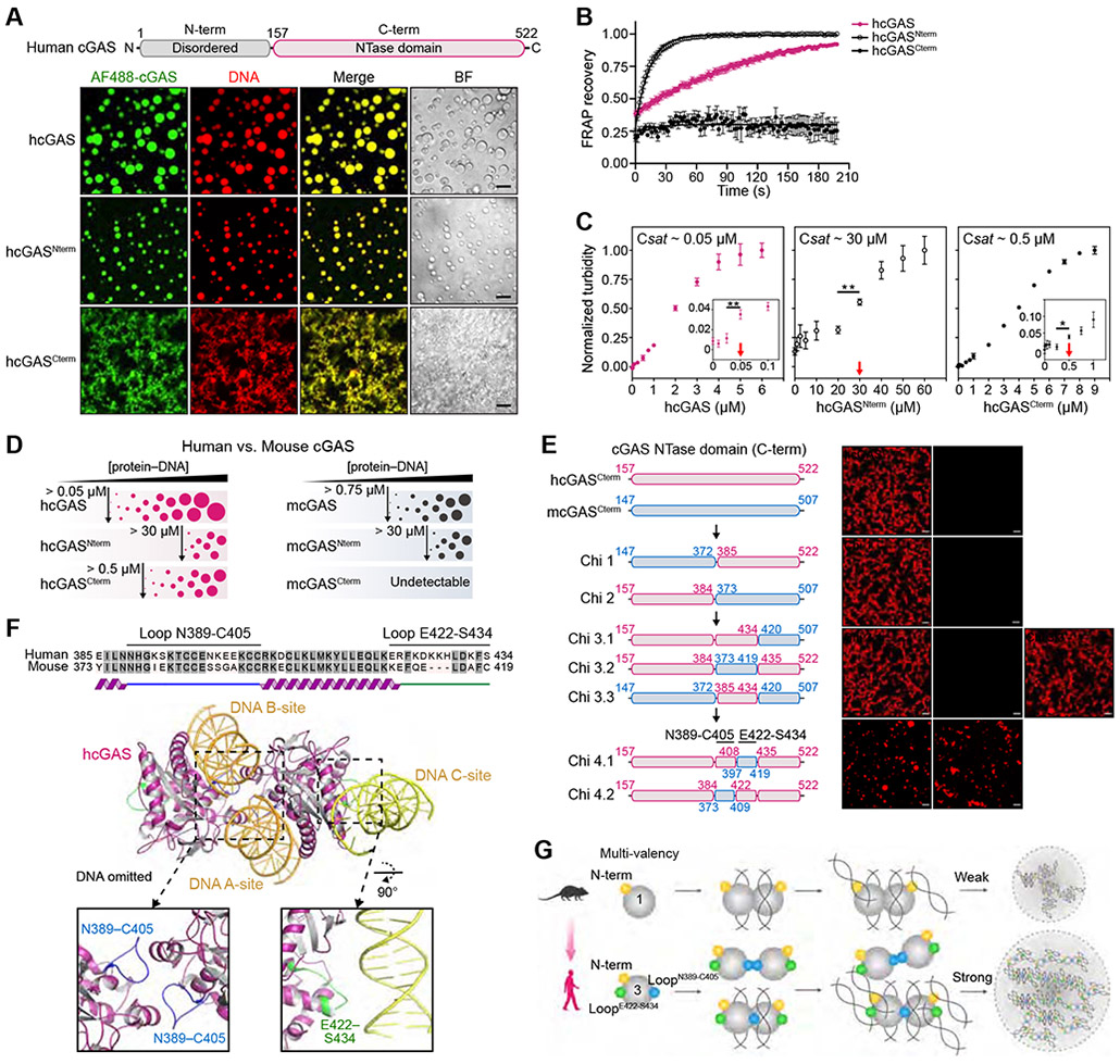
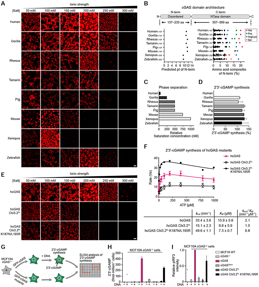
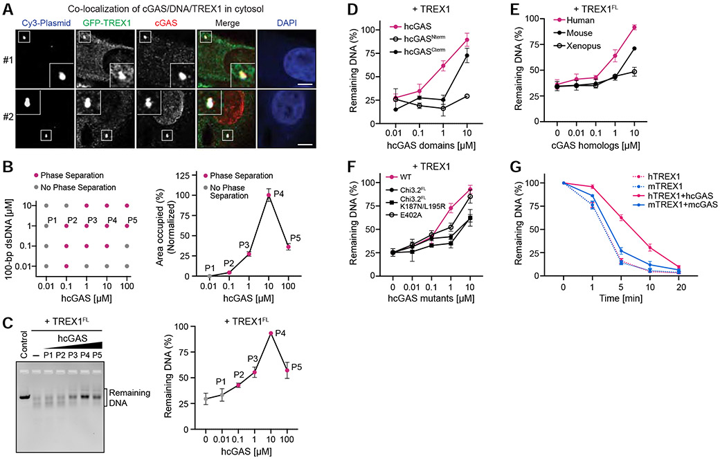
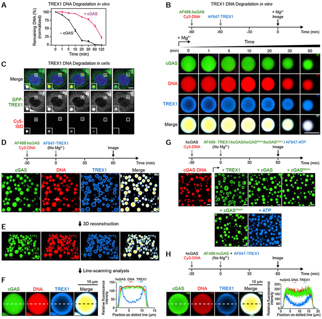
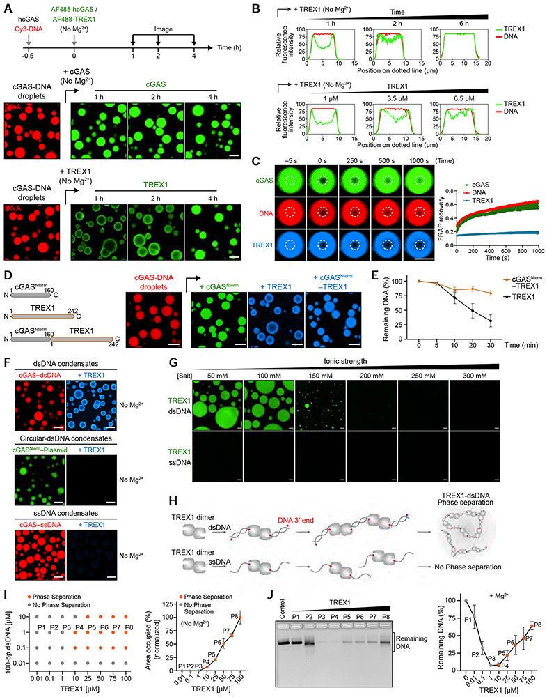
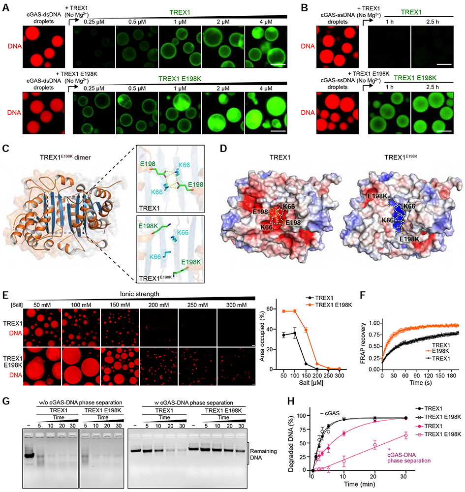
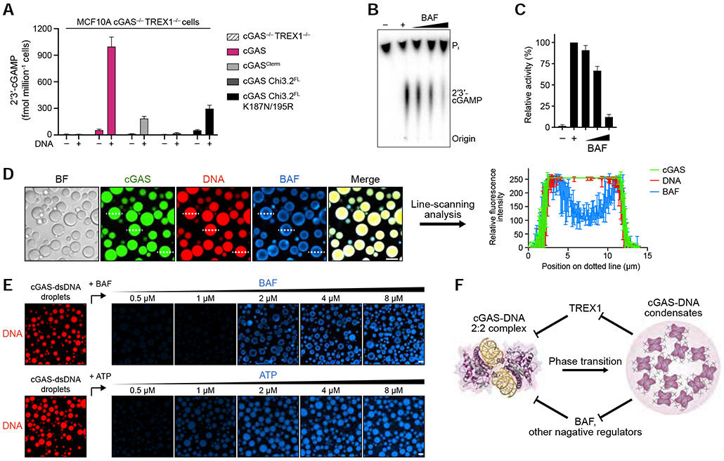
Cyclic GMP-AMP synthase (cGAS) recognition of cytosolic DNA is critical for the immune response to cancer and pathogen infection. Here, we discover that cGAS-DNA phase separation is required to resist negative regulation and allow efficient sensing of immunostimulatory DNA. We map the molecular determinants of cGAS condensate formation and demonstrate that phase separation functions to limit activity of the cytosolic exonuclease TREX1. Mechanistically, phase separation forms a selective environment that suppresses TREX1 catalytic function and restricts DNA degradation to an outer shell at the droplet periphery. We identify a TREX1 mutation associated with the severe autoimmune disease Aicardi-Goutières syndrome that increases penetration of TREX1 into the repressive droplet interior and specifically impairs degradation of phase-separated DNA. Our results define a critical function of cGAS-DNA phase separation and reveal a molecular mechanism that balances cytosolic DNA degradation and innate immune activation.

Keywords: TREX1; cGAS; innate immunity; phase separation.


Conflict of interest statement

Declaration of interests The authors declare no competing interests.

Figures

Figure 1.
Figure 1.. Molecular mechanism of cGAS-DNA phase separation
(A) Top, schematic of the domain architecture of human cGAS (hcGAS). Bottom, fluorescence microscopy of DNA-induced phase separation of hcGAS proteins with bright field (BF) images. Recombinant hcGAS (10 μM), hcGASNterm (80 μM), and hcGASCterm (20 μM) were incubated with 100 bp dsDNA (10 μM) in buffer containing 250 mM (hcGAS), 150 mM (hcGASNterm), or 250 mM salt (hcGASCterm). Scale bar, 10 μm. (B) FRAP analysis of cGAS-DNA phase-separated condensates. Time 0 indicates the time of photobleaching. Data represent the mean ± SEM of 7 droplets in 3 independent experiments. (C) Analysis of relative saturation concentrations of cGAS proteins by turbidity assay. A series of concentrations of protein with 100 bp dsDNA (equal amounts) were mixed at 150 mM salt, and the absorbance of 340 nm was used as the readout of turbidity. The relative saturation concentrations are indicated with red arrows. All data are expressed as the mean ± SEM of more than 5 independent experiments. Statistical significance was calculated with a two-tailed T test, **p = <0.01, *p = <0.1. (D) Schematic representing the phase behaviors and saturation concentrations of hcGAS and mouse cGAS (mcGAS) proteins determined by fluorescence intensity as in Figures S1F,G and S2C,D. (E) Chimera experiments mapping the molecular determinant of enhanced human cGAS phase separation to two loops hcGAS N389–C405 and E422–S434. Left, schematic of cGAS constructs. Amino acid numbers are colored magenta for hcGAS and blue for mcGAS. Right, fluorescence microscopy images analyzing phase separation. cGAS chimeras (10 μM) were incubated with 100 bp dsDNA (10 μM) at 150 mM salt. Scale bar, 10 μm. (F) Structure of the hcGAS–DNA complex (derived from combining PDB 6CT9 and additional DNA from PDB 6EDB) and schematic highlight of the mapped hcGAS loop sequences required for enhancement of cGAS–DNA phase separation. (G) Cartoon model of the molecular basis of cGAS-DNA phase separation. See also Figures S1, S2, and Video S1.
Figure 2.
Figure 2.. cGAS phase separation does not directly control 2′3′-cGAMP synthesis in vitro but is required for immune activation in cells
(A) Fluorescence microscopy images of DNA-induced phase separation of cGAS homologs. Phase separation was induced as in Figure 1A with recombinant cGAS homologs and 100 bp dsDNA in buffer with varying salt concentration. Scale bars, 10 μm. (B) Schematic of cGAS domain architecture along with the predicted isoelectric point (pI) and amino acid composition of the N-terminal regions. (C) Relative saturation concentrations of each cGAS homolog required for phase separation determined by turbidity assay. (D) In vitro 2′3′-cGAMP synthesis activity of each cGAS homolog (see STAR Methods). Data were normalized to hcGAS as 100% and represent the mean ± SEM of 3 independent experiments. (E) Fluorescence microscopy images of DNA-induced phase separation of hcGAS phase separation mutants. Phase separation was induced as in Figure 1A. Scale bars, 10 μm. (F) Analysis of hcGAS and phase separation mutant enzyme kinetics. cGAS protein enzyme activity was measured as function of varying ATP concentration, and 2′3′-cGAMP synthesis was quantified and fit according to the Michaelis-Menten kinetics. Data represent the mean ± SEM in 3 independent experiments. (G) Schematic of analysis of cGAS activity in cells (see STAR Methods). (H,I) ELISA analysis of 2′3′-cGAMP production and quantification of immunoblot analysis of IRF3 phosphorylation in MCF10A cGAS−/− cells reconstituted with hcGAS or hcGAS phase separation mutant alleles and transfected with plasmid DNA. Data are the mean ± SD of 3 experiments. See also Figure S3.
Figure 3.
Figure 3.. cGAS phase separation resists DNA degradation by exonuclease TREX1
(A) Imaging of co-localization of cGAS/DNA/TREX1 in the cell cytosol (see STAR Methods). cGAS staining indicates that cGAS predominantly localizes in either the cytosol (#1) or nucleus (#2). Scale bar, 5 μm. (B) Left, phase diagram of purified hcGAS under various protein and DNA concentrations at 150 mM salt, where grey dots indicate no phase separation, and magenta dots indicate observable condensate formation (see also C). Right, quantification of phase separation with 1 μM DNA and increasing concentration of hcGAS. Data represent the percent area of the image occupied by condensates relative to the maximal phase separation observed with 10 μM hcGAS and are plotted as the mean ± SEM of 17 images from 3 independent experiments. (C) cGAS phase separation protects DNA from TREX1 degradation. Left, in vitro analysis of DNA degradation by full-length TREX1 (TREX1FL) in the presence of varying degrees of cGAS-DNA phase separation. Phase separation was induced with increasing cGAS concentration as shown in (B) in the presence of 1 μM 100-bp dsDNA, and DNA degradation was measured by incubating with purified TREX1FL and resolving the remaining DNA on an agarose gel. Right, quantification of remaining DNA. Data represent the mean ± SEM of 5 independent experiments. (D) Analysis of the TREX1 resistance by individual cGAS domains. Phase separation was induced with 1 μM DNA and titrations of N-term and C-term cGAS at indicated concentrations. DNA degradation was then initiated by adding TREX1 (M1–K242) at 0.1 μM. Remaining DNA was quantified as in (C). Data are represented as the mean ± SEM of 3 independent experiments. (E) Analysis of TREX1 resistance by cGAS homologs. Phase separation was induced with 1 μM DNA and a titration of cGAS homolog protein as indicated. DNA degradation was measured as in (C). Data represent the mean ± SEM of 3 independent experiments. (F) Inhibition of cGAS-DNA phase separation reduces TREX1 resistance. DNA degradation by TREX1 was measured in the presence of hcGAS or hcGAS mutants. Experiments were performed and data were quantified as in (C). Data are represented as the mean ± SEM of 4 independent experiments. (G) Analysis of species-specific TREX1 resistance by cGAS-DNA phase separation. Human and mouse TREX1 (hTREX1 and mTREX1) DNase activity was assessed in the absence or presence of cGAS phase separation as indicated. Remaining DNA was resolved on an agarose gel and quantified. Data represent the mean ± SEM of 4 independent experiments. See also Figure S4.
Figure 4.
Figure 4.. cGAS phase separation restricts TREX1 DNA degradation to an outer shell at the droplet periphery
(A) Comparison of TREX1 DNA degradation activity with (black) and without (magenta) cGAS-DNA phase separation at 250 mM salt. Time 0 indicates addition of 5 mM Mg2+ to activate TREX1. Data represent the mean ± SEM of 3 independent experiments. (B) Time-lapse imaging of TREX1 degradation of cGAS-DNA droplets in vitro. Droplet formation was induced by hcGAS (10 μM), 100-bp DNA (10 μM) and TREX1 (1 μM) as indicated. Concentrations of cGAS and TREX1 are based on the cellular cGAS concentration being 10-fold higher than TREX1 (Hein et al., 2015). Time 0 indicates addition of 5 mM MgCl2 to activate TREX1. Scale bar, 10 μm. (C) Time-lapse imaging of TREX1 degradation of immunostimulatory DNA (ISD) puncta in cells (see STAR Methods). Time 0 represents the start of imaging 2 h after initial Cy5-ISD transfection. Scale bar, 5 μm. (D) Fluorescence microscopy images showing a shell-like formation of TREX1 around cGAS-DNA droplets at the early stage of degradation. cGAS-DNA-TREX1 droplet formation was induced as in (B) without adding MgCl2. The droplet labeled with a dotted line was selected for further line-scanning analysis in (F). Scale bar, 10 μm. (E,F) Three-dimensional reconstruction and line-scanning analysis of cGAS-DNA-TREX1 droplets in (D). Corresponding fluorescence intensity along the dotted line is shown to the right. (G) Fluorescence microscopy images showing cGAS-DNA droplets inhibit TREX1 access but not cGAS incorporation. cGAS-DNA droplet formation was induced as illustrated above. Fluorescently labeled components as indicated were subsequently added and incubated for additional time as indicated. Scale bar, 10 μm. (H) cGAS-DNA droplet formation selectively resists TREX1 incorporation. hcGAS-DNA droplet formation was induced using non-labeled hcGAS and Cy3-labeled DNA, followed by simultaneously adding 1 μM AF647-labeled TREX1 and 1 μM AF488-labeled hcGAS. Images were collected after 1 h and fluorescence intensity along the dotted line is shown to the right. All imaging data are representative of at least 3 independent experiments. See also Figure S5, and Videos S2 and S3.
Figure 5.
Figure 5.. Phase separation is a direct suppressor of TREX1 exonuclease activity
(A) Time-lapse imaging of slower partitioning of TREX1 into cGAS-DNA droplets compared to cGAS. cGAS-DNA droplets were preformed as indicated, and the incorporation rates of TREX1 and cGAS were tracked using fluorescently labeled components. Scale bar, 10 μm. (B) Line-scanning analysis of TREX1 incorporation demonstrates that TREX1 migration to the center of cGAS-DNA droplets requires extended incubation times (top) or elevated TREX1 protein levels (bottom). (C) Analysis of the fluidity of TREX1, cGAS and DNA in cGAS-DNA droplets. cGAS-DNA droplets were incubated for 4 h to allow complete incorporation of TREX1 and then analyzed by FRAP to measure component fluidity. Time 0 indicates the time of photo bleaching. Plots are generated from 12 droplets and data represent the mean ± SEM of 3 independent experiments. (D) Schematic of engineered cGAS-TREX1 fusion constructs developed to permit rapid incorporation of TREX1 into cGAS-DNA droplets (Left). Right, fluorescence microscopy images of construct incorporation into preformed cGAS-DNA droplets as in Figure 4G. Scale bar, 10 μm. (E) Entry into cGAS-DNA droplets restricts TREX1 nuclease activity. Following induction of cGAS-DNA phase separation, DNA degradation was initiated with addition of TREX1 or cGASNterm–TREX1 and remaining DNA was quantified by agarose gel analysis. Data represent the mean ± SEM of 3 independent experiments. (F) Fluorescence microscopy images of TREX1 interactions with cGAS-DNA droplets formed with linear dsDNA, closed circular dsDNA, or ssDNA. Scale bar, 10 μm. (G) Fluorescence microscopy images showing dsDNA-induced phase separation of TREX1. TREX1-dsDNA droplet formation was induced by TREX1 (20 μM) and 100-bp dsDNA (10 μM) at various salt concentrations as indicated. Scale bar, 10 μm. (H) Schematic of hypothetical TREX1-dsDNA interactions that drive liquid-liquid phase separation. (I) Left, phase diagram of purified TREX1 under various protein and dsDNA concentrations at 150 mM salt with no MgCl2. Dots indicate conditions where a single phase (grey) or two phases (orange) are present. Right, quantification of TREX1-dsDNA phase separation with 1 μM dsDNA and increasing concentration of TREX1. Data correspond to the percent area of the image occupied by droplets and were quantified relative to maximal phase separation observed with 100 μM TREX1. Data represent the mean ± SEM of 7 images in 3 independent experiments. (J) TREX1-dsDNA phase separation inhibits TREX1 exonuclease activity. TREX1-dsDNA phase separation was induced with 100-bp dsDNA and increasing TREX1 concentration as shown in (I) in the presence of 5 mM MgCl2. Reactions were inactivated and DNA was quantified by agarose gel analysis. Data represent the mean ± SEM of 3 independent experiments. See also Figures S5, S6, and Video S4.
Figure 6.
Figure 6.. The disease mutation TREX1 E198K alters interactions with cGAS-DNA droplets
(A) Fluorescence microscopy images of incorporation of TREX1 and TREX1 E198K into cGAS-DNA droplets. cGAS-dsDNA droplet formation was induced as in Figure 4G, and TREX1-incorporation was analyzed with increasing concentration of AF488-labeled TREX1 (top) and TREX1 E198K (bottom). Scale bar, 10 μm. (B) Time-lapse images of incorporation of TREX1 into cGAS-ssDNA droplets. cGAS-ssDNA droplets were formed as in Figure 5F, and AF488-labeled TREX1 (top) or TREX1 E198K (bottom) was added to the preformed ssDNA droplets. Scale bar, 10 μm. (C) 1.8 Å crystal structure of the mouse TREX1E198K dimer with zoom-in cutaways of the locations of residues E198 and K66 in wildtype mTREX1 (top, PDB: 3MXJ) and mTREX1 E198K (bottom). The 2FO–FC electron density map is contoured at 1.0 σ for TREX1 and 0.7 σ for TREX1E198K (D) The crystal structure of TREX1E198K reveals extensive remodeling of surface electrostatic potential. Surface electrostatic potentials of TREX1 and TREX1 E198K (blue positive, red negative) with E198, K66, and E198K positions highlighted with dashed outlines. (E) Left, fluorescence microscopy images of TREX1-dsDNA (top) and TREX1E198K-dsDNA (bottom) phase separation with various salt concentrations. Scale bar, 10 μm. Right, quantification of phase separation by the percent area of the image occupied by droplets. Data are represented as the mean ± SEM of 9 images in 3 independent experiments. (F) FRAP analysis of TREX1-dsDNA (black) and TREX1E198K-dsDNA (orange) droplets formed by mixing 100-bp dsDNA and unlabeled TREX1 or TREX1E198K protein for 3 h. Time 0 indicates the time of photobleaching pulse. Plots are generated from 6 droplets and data represent the mean ± SEM of 3 independent experiments. (G,H) In vitro analysis and quantification of exonuclease activity of TREX1 mutation E198K in the absence or presence of cGAS phase separation. cGAS-DNA phase separation was induced as in Figure 5E, and DNA degradation initiated by adding TREX1 and TREX1 E198K. Remaining DNA was resolved on an agarose gel and quantified. Data represent the mean ± SEM of 3 independent experiments. See also Figure S7 and Video S5.
Figure 7.
Figure 7.. cGAS phase separation resists immune suppression by multiple negative regulators
(A) ELISA analysis of 2′3′-cGAMP production in MCF10A cGAS−/− TREX1−/− cells reconstituted with hcGAS or hcGAS mutant alleles and transfected with plasmid DNA. Data are the mean ± SD of 3 experiments. (B,C) In vitro analysis of BAF-dependent inhibition of cGAS 2′3′-cGAMP synthesis. Purified hcGAS enzyme was stimulated with 100-bp DNA in reactions supplemented with an increasing concentration of BAF and 2′3′-cGAMP production was analyzed and quantified as in Figure S1B. Data represent the mean ± SEM of 4 independent experiments. (D) Fluorescence microscopy images (left) and line-scanning analysis (right) showing that BAF is excluded to an outer shell at the cGAS-DNA droplet periphery. cGAS-DNA droplet formation was induced as in Figure 4D. Scale bar, 10 μm. Fluorescence intensity along the dotted lines was analyzed (right). Plots are generated from 3 droplets and data represent the mean ± SD. (E) Fluorescence microscopy images of dosage-dependent incorporation of BAF and ATP into cGAS-DNA droplets. cGAS-dsDNA droplet formation was induced as in Figure 5A, and BAF/ATP incorporation was analyzed with increasing concentration of AF647-labeled BAF (top) or AF647-labeled ATP (bottom). Scale bar, 10 μm. (F) Model of the role of cGAS-DNA phase separation in resisting negative regulation. DNA-induced cGAS phase separation creates a selective environment that suppresses entry of negative regulators and allows sensing of immunostimulatory DNA. See also Figure S7.

Comment in

References

    1. Ablasser A, and Chen ZJ (2019). cGAS in action: Expanding roles in immunity and inflammation. Science 363, eaat8657. - PubMed
    1. Ablasser A, and Hur S (2020). Regulation of cGAS- and RLR-mediated immunity to nucleic acids. Nat. Immunol 21, 17–29. - PubMed
    1. Ablasser A, Goldeck M, Cavlar T, Deimling T, Witte G, Röhl I, Hopfner K-P, Ludwig J, and Hornung V (2013). cGAS produces a 2′–5′–linked cyclic dinucleotide second messenger that activates STING. Nature 498, 380–384. - PMC - PubMed
    1. Adams PD, Afonine PV, Bunkóczi G, Chen VB, Davis IW, Echols N, Headd JJ, Hung L-W, Kapral GJ, Grosse-Kunstleve RW, et al. (2010). PHENIX: a comprehensive Python-based system for macromolecular structure solution. Acta Crystallogr D Biol Crystallogr 66, 213–221. - PMC - PubMed
    1. Alshareedah I, Moosa MM, Raju M, Potoyan DA, and Banerjee PR (2020). Phase transition of RNA-protein complexes into ordered hollow condensates. Proceedings of the National Academy of Sciences 117, 15650–15658. - PMC - PubMed

Publication types

MeSH terms

Substances

Supplementary concepts