. 2021 Mar 9;16(3):566-581.
doi: 10.1016/j.stemcr.2021.01.009. Epub 2021 Feb 18.

Rbbp4 Suppresses Premature Differentiation of Embryonic Stem Cells

Affiliations

Rbbp4 Suppresses Premature Differentiation of Embryonic Stem Cells

Yikai Huang et al. Stem Cell Reports. .

Abstract

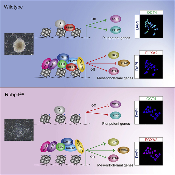
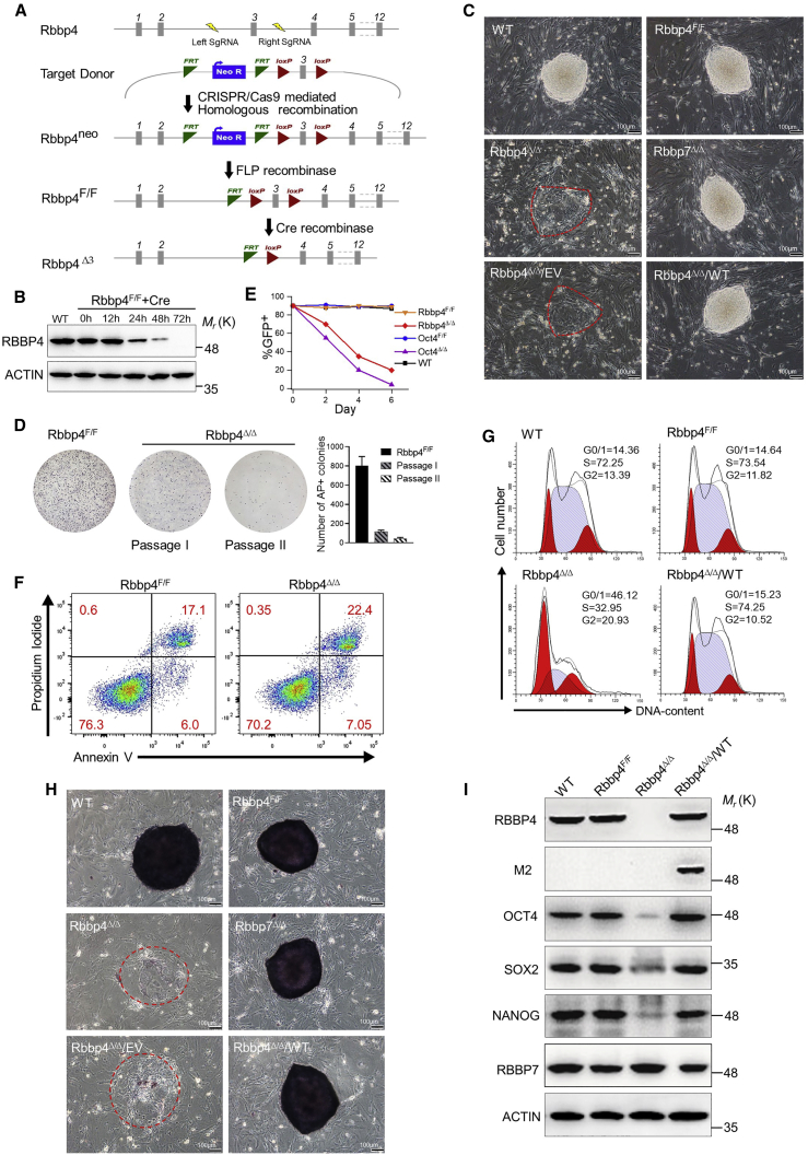
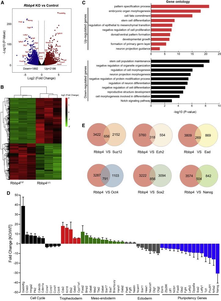
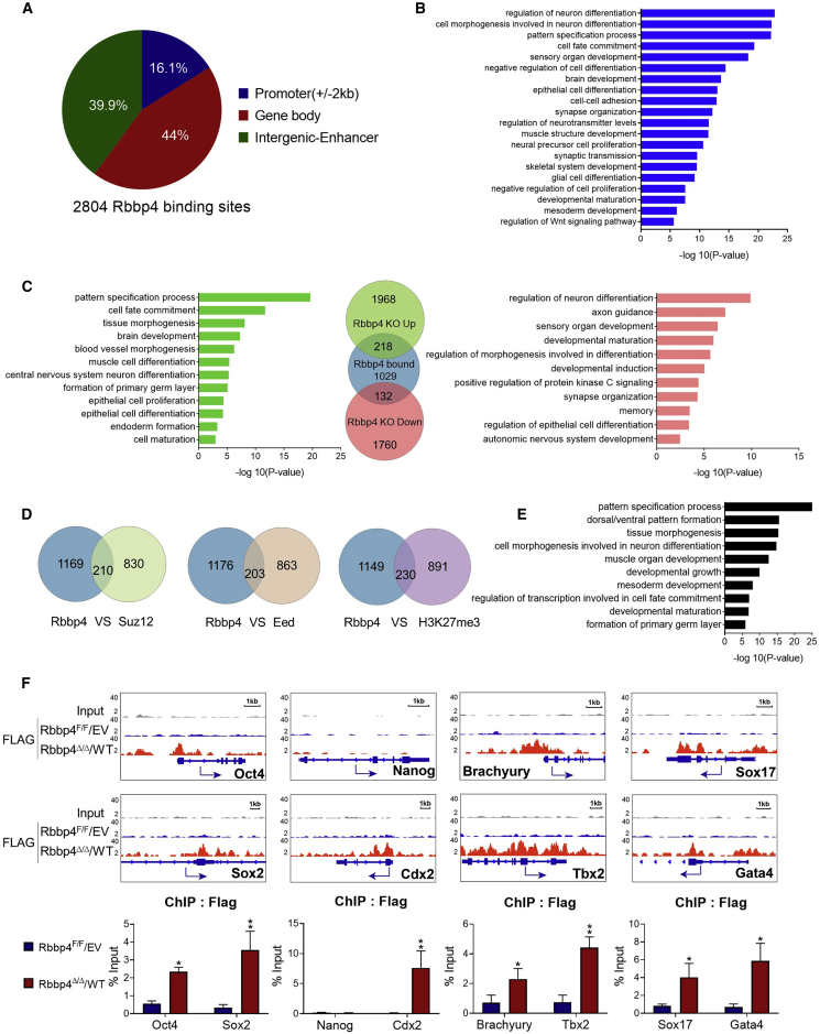
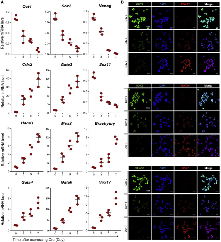
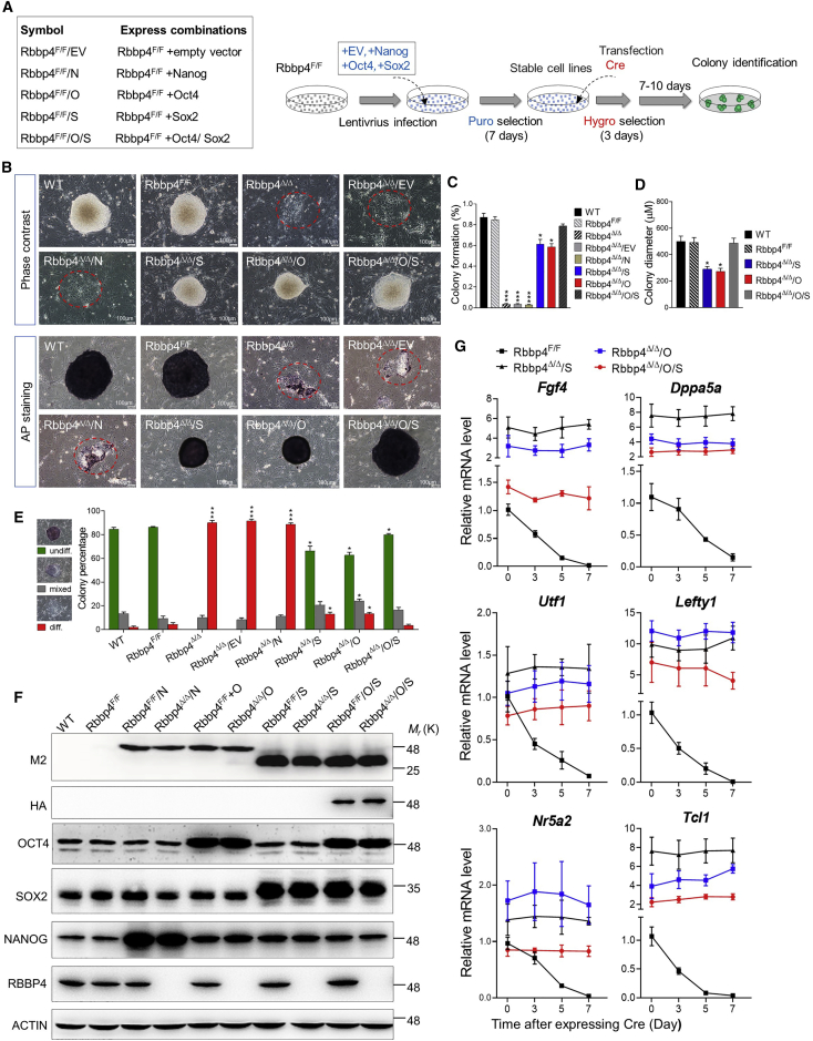
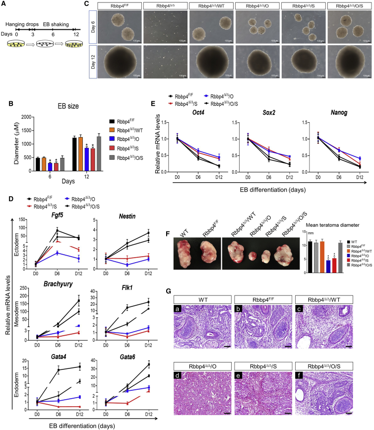
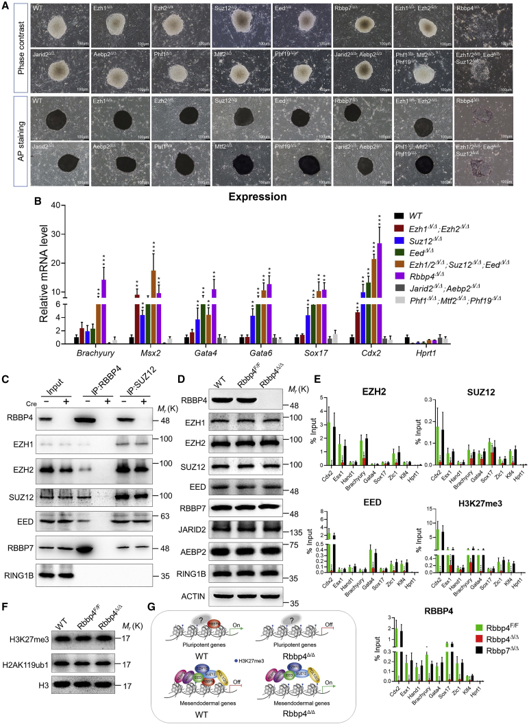
Polycomb group (PcG) proteins exist in distinct multi-protein complexes and play a central role in silencing developmental genes, yet the underlying mechanisms remain elusive. Here, we show that deficiency of retinoblastoma binding protein 4 (RBBP4), a component of the Polycomb repressive complex 2 (PRC2), in embryonic stem cells (ESCs) leads to spontaneous differentiation into mesendodermal lineages. We further show that Rbbp4 and core PRC2 share an important number of common genomic targets, encoding regulators involved in early germ layer specification. Moreover, we find that Rbbp4 is absolutely essential for genomic targeting of PRC2 to a subset of developmental genes. Interestingly, we demonstrate that Rbbp4 is necessary for sustaining the expression of Oct4 and Sox2 and that the forced co-expression of Oct4 and Sox2 fully rescues the pluripotency of Rbbp4-null ESCs. Therefore, our study indicates that Rbbp4 links maintenance of the pluripotency regulatory network with repression of mesendoderm lineages.

Keywords: Oct4; PRC2; RBBP4; Sox2; embryonic stem cells; mesendoderm; pluripotency; polycomb; self-renewal.


Figures

None
Graphical abstract
Figure 1
Figure 1
Rbbp4 is essential in the maintenance of self-renewal and pluripotency in ESCs (A) Schematic representation of the production for conditional inactivation of Rbbp4 in ESCs. (B) Western blot showing RBBP4 levels in Rbbp4F/F-transfected Cre recombinase for different time points. (C) Morphology of ESC colonies of indicated genotypes. Bright-field images of ESC colonies after 7 days of culture (grown from a single ESC). All ESC colony images were photographed at day 7 after seeding single-cell suspensions on feeder layers. Scale bar, 100 μm. The outlines of obviously defective colonies were circled in red. Rbbp4Δ/Δ ESCs expressing empty FLAG vector (Rbbp4Δ/Δ/EV) and vectors encoding wild-type RBBP4-FLAG (Rbbp4Δ/Δ/WT) are also shown. (D) Secondary ES colony-replating assay. Bar graph shows the number of cells with AP-positive staining in the absence of RBBP4. Error bars represent means and STD from three 6 cm dishes. (E) ESC growth competition assay. Oct4 knockout ESCs serve as a negative control. (F) Representative fluorescence-activated cell sorting plots of annexin V and propidium iodide (PI) levels in Rbbp4F/F and Rbbp4Δ/Δ ESCs. Percentages of cells with different apoptosis marker levels are shown. (G) Cell-cycle analysis of indicated ESCs. Percentages of cells in different phases are indicated. Representative histograms presented here show distribution of cells in sequential phases (G0/G1, red; S, light blue; and G2/M, red) of cell cycle. (H) Alkaline phosphatase activity staining on each indicated ESC colony. Scale bar, 100 μm. (I) Western blot analysis of OCT4, SOX2, NANOG, RBBP4, and RBBP7 protein expression in indicated ESCs; ACTIN served as a loading control. See also Figures S1 and S2.
Figure 2
Figure 2
Loss of Rbbp4 results in aberrant expression of ESC core pluripotency factors and differentiation-associated genes (A) Volcano plot showing the distribution of the differentially expressed (DE) genes with 2-fold changes upon Rbbp4 deletion. p < 0.05. Up- and downregulated genes are colored red and blue, respectively. (B) Heatmap illustrating the RNA expression in Rbbp4F/F and Rbbp4Δ/Δ ESCs of RNA-seq analysis for 2-fold expression differentially expressed genes. False discovery rate < 0.05. Up- and downregulated genes are reported as red and green, respectively. (C) Gene ontology (GO) enrichment analyses for biological processes associated with genes differentially expressed upon Rbbp4 deletion in ESCs. Analysis was carried out using Metascape (Zhou et al., 2019). (D) Validation of RNA-seq data by qRT-PCR analysis. Relative mRNA levels of indicated cell-cycle-related genes, lineage-specific genes, and pluripotency-related genes in Rbbp4F/F after 3 days of Cre transfection were measured and data were normalized to β-actin relative to Rbbp4F/F. Data are pooled from three independent experiments and the error bars represent standard deviation of triplicate qPCR data. (E) Venn diagrams (top) showing the overlap of the target genes between Rbbp4 and the core subunits of the PRC2 complex, respectively (Das et al., 2015; Pasini et al., 2007). Venn diagrams (bottom) showing the numbers of the regulated genes between Rbbp4 and pluripotency markers (Oct4, Sox2, and Nanog) (Ding et al., 2015; Loh et al., 2006). See also Figure S3.
Figure 3
Figure 3
RBBP4 shares target genes with PRC2 (A) Pie chart showing the distribution of RBBP4 binding sites in mouse ESCs. (B) GO enrichment analyses for genes that RBBP4 binds to. The biological processes of the top 20 are shown. (C) Venn diagram showing the overlap between genes differentially expressed after Rbbp4 deletion and those occupied by RBBP4. GO analysis for biological processes associated with the overlapping genes. (D) Venn diagram showing the overlap between genes that RBBP4 binds to and those occupied by SUZ12, EED, or H3K27me3. (E) GO analysis of overlapping genes between RBBP4 binds and PRC2 targets. (F) Genome browser tracks to show RBBP4 occupancy near lineage-specific markers for trophectoderm (Cdx2), mesoderm (Brachyury, Tbx2), and endoderm (Sox17, Gata4), Oct4 and Sox2 (up). ChIP-qPCR data showing binding of RBBP4-FLAG to representative RBBP4 target promoters (bottom). Data are plotted as mean ± SD. (n = 3). p < 0.05, ∗∗p < 0.01. See also Figure S4.
Figure 4
Figure 4
Rbbp4 knockout significantly reduces the expression of pluripotency-associated genes and promotes mesendodermal gene expression in ESCs (A) Time-course analyses of pluripotency-associated genes and the lineage-specific markers for trophectoderm, mesoderm, and endoderm expression levels in Rbbp4F/F ESCs after transient expression of Cre recombinase. Expression is normalized by β-actin. (B) Immunofluorescence showing co-staining of the pluripotency markers (OCT4, SOX2, and NANOG) and the lineage-specific markers (FOXA2, GATA6, and CDX2) at the indicated time points after expressing Cre recombinase in Rbbp4F/F ESCs, respectively. DAPI (blue), pluripotency marker genes (green), the lineage-specific markers (red), and the merge picture are shown. Magnification, 63×. Data are represented as mean ± SD of three independent experiments.
Figure 5
Figure 5
Rbbp4 governs pluripotency by maintaining the expression of Oct4 and Sox2 (A) Left: reference legend for cell lines used in Figure 5. Right: experimental diagrams of rescue assay. (B) Representative images of the ESC colony of the indicated genotypes cultured for 7 days. Top, phase-contrast microscopy; bottom, AP staining. (C) Percentage of isolated single ESCs of the indicated genotypes giving rise to macroscopic colonies. (D) Bar graphs show the mean diameter of 30 random ESC colonies of the indicated genotypes. (E) Quantitative analysis of colony formation assay in ESCs of indicated genotypes. AP-stained colonies were scored as undifferentiated (undiff.), mixed or differentiated (diff.). (F) Western blot analysis of OCT4, SOX2, and NANOG protein levels after the overexpression of Oct4, Sox2, and Nanog in Rbbp4F/F ESCs and Rbbp4Δ/Δ ESCs. (G) Time-course analysis of pluripotency-associated gene expression after expressing Cre in the corresponding cell lines by qRT-PCR. Data in (C–E) represent the mean ± SD of three independent experiments. p < 0.05, ∗∗p < 0.01, and ∗∗∗p < 0.001 (Student's t test) compared with the control.
Figure 6
Figure 6
Overexpression of Sox2 and Oct4 maintains ESC self-renewal and pluripotency in the absence of Rbbp4 in vitro and in vivo (A) Schematic illustration of the embryoid body (EB) formation procedure for differentiation of ESCs. (B) The diameter statistics of EB. Thirty 30 EB diameters of each genotype were measured at each time point. (C) Phase-contrast images of floating EB derived from indicated ESCs at day 6 and day 12. Scale bar, 100 μm. (D) qRT-PCR analysis of lineage-specific markers at day 0, day 6, and day 12 during EB formation from the indicated ESC lines. Fgf5, Nestin (ectoderm), Brachyury, Flk1 (mesoderm), and Gata4, Gata6 (endoderm). Data are normalized to the expression levels in ESCs. Error bars represent ± SD (n = 3). (E) qRT-PCR mRNA analysis of pluripotency markers during time-course differentiation of Rbbp4F/F, and rescue of Rbbp4 in Rbbp4Δ/Δ EBs. Samples were collected at different days. (F) In vivo differentiation potential of formation teratomas of indicated ESCs. Left: the morphological characteristics of teratomas are shown. Right: average teratoma size at 4 weeks after injection in the indicated groups. (G) Histological analysis of teratomas. (A–F) Histological sections of hematoxylin and eosin-stained teratomas. (A, B, C, F) Each teratoma contained three embryonic germ layer tissues. (D and E) Immature germ layer differentiation is shown. Scale bar, 50 μm. Data in (B and D–F) represent mean ± SD obtained from three independent experiments. p < 0.05 and ∗∗p < 0.01 (Student's t test) compared with the control.
Figure 7
Figure 7
RBBP4 guides PRC2 recruitment and H3K27 trimethylation at genomic loci (A) Bright-field images of an ESC colony of wild-type and indicated genotypes after 7 days of culture. AP staining images of the ESC colony that arose from the wild-type and indicated genotypes. Scale bar, 100 μm. (B) qRT-PCR quantification of Rbbp4 targets in indicated cell lines normalized to β-actin. Hprt1 is represented as a negative control. (C) Endogenous coimmunoprecipitations of RBBP4 and SUZ12 in Rbbp4F/F transfected with Cre (+) or control vector (−) for 72 h, followed by western blot analysis with the indicated antibodies against core subunits of PRC2. (D) Western blot analyses using the indicated antibodies against PRC2 subunits on whole-cell lysates from wild-type (WT), Rbbp4F/F, and Rbbp4Δ/Δ ESCs. (E) ChIP-qPCR analysis of EZH2, SUZ12, EED, H3K27me3, and RBBP4 binding at the indicated regions of pluripotent transcription factors and mesendodermal genes (normalized to input) in Rbbp4F/F, Rbbp7Δ/Δ, or Rbbp4F/F ESCs that were transfected with Cre after 3 days. (F) Western blot for H3K27 tri-methylation and H2AK119 mono-ubiquitination on nuclear lysates from Rbbp4Δ/Δ and matched control ESCs. Histone 3 was used as a loading control. (G) Proposed model of Rbbp4-mediated pluripotency maintenance of ESCs. Data in (B and E) represent the mean ± SD of three independent experiments. p < 0.05, ∗∗p < 0.01, and ∗∗∗p < 0.001 (Student's t test) compared with the control. See also Figures S5 and S6.

Similar articles

Cited by

References

    1. Blackledge N.P., Farcas A.M., Kondo T., King H.W., McGouran J.F., Hanssen L.L., Ito S., Cooper S., Kondo K., Koseki Y. Variant PRC1 complex-dependent H2A ubiquitylation drives PRC2 recruitment and polycomb domain formation. Cell. 2014;157:1445–1459. - PMC - PubMed
    1. Boyer L.A., Lee T.I., Cole M.F., Johnstone S.E., Levine S.S., Zucker J.P., Guenther M.G., Kumar R.M., Murray H.L., Jenner R.G. Core transcriptional regulatory circuitry in human embryonic stem cells. Cell. 2005;122:947–956. - PMC - PubMed
    1. Chamberlain S.J., Yee D., Magnuson T. Polycomb repressive complex 2 is dispensable for maintenance of embryonic stem cell pluripotency. Stem Cells. 2008;26:1496–1505. - PMC - PubMed
    1. Chetty S., Pagliuca F.W., Honore C., Kweudjeu A., Rezania A., Melton D.A. A simple tool to improve pluripotent stem cell differentiation. Nat. Methods. 2013;10:553–556. - PMC - PubMed
    1. Conklin J.F., Baker J., Sage J. The RB family is required for the self-renewal and survival of human embryonic stem cells. Nat. Commun. 2012;3:1244. - PubMed

Publication types