. 2021 May 21:517:110630.
doi: 10.1016/j.jtbi.2021.110630. Epub 2021 Feb 17.

Structure-function relationships in the feto-placental circulation from in silico interpretation of micro-CT vascular structures

Affiliations

Structure-function relationships in the feto-placental circulation from in silico interpretation of micro-CT vascular structures

Monika Byrne et al. J Theor Biol. .

Abstract

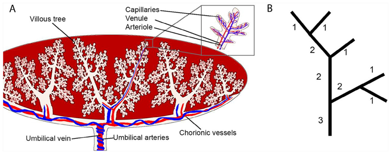
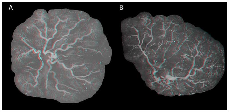
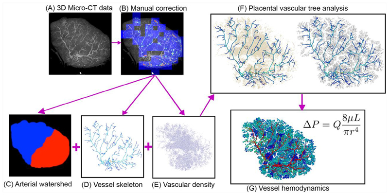
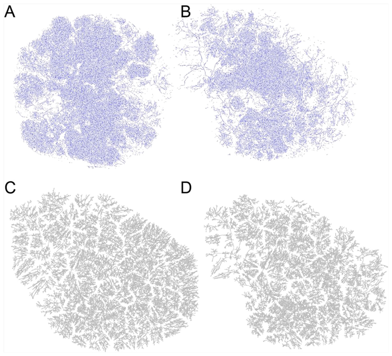
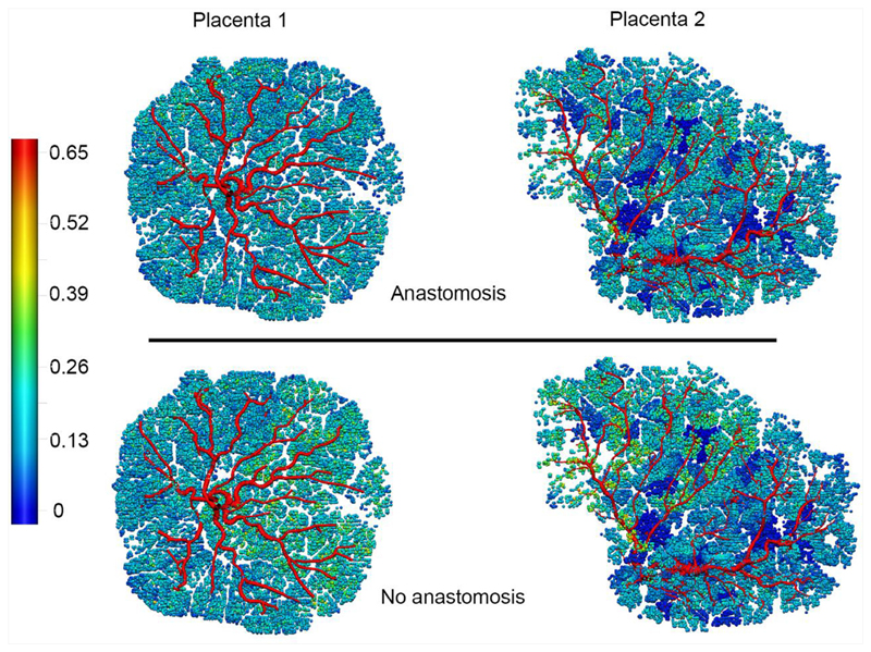
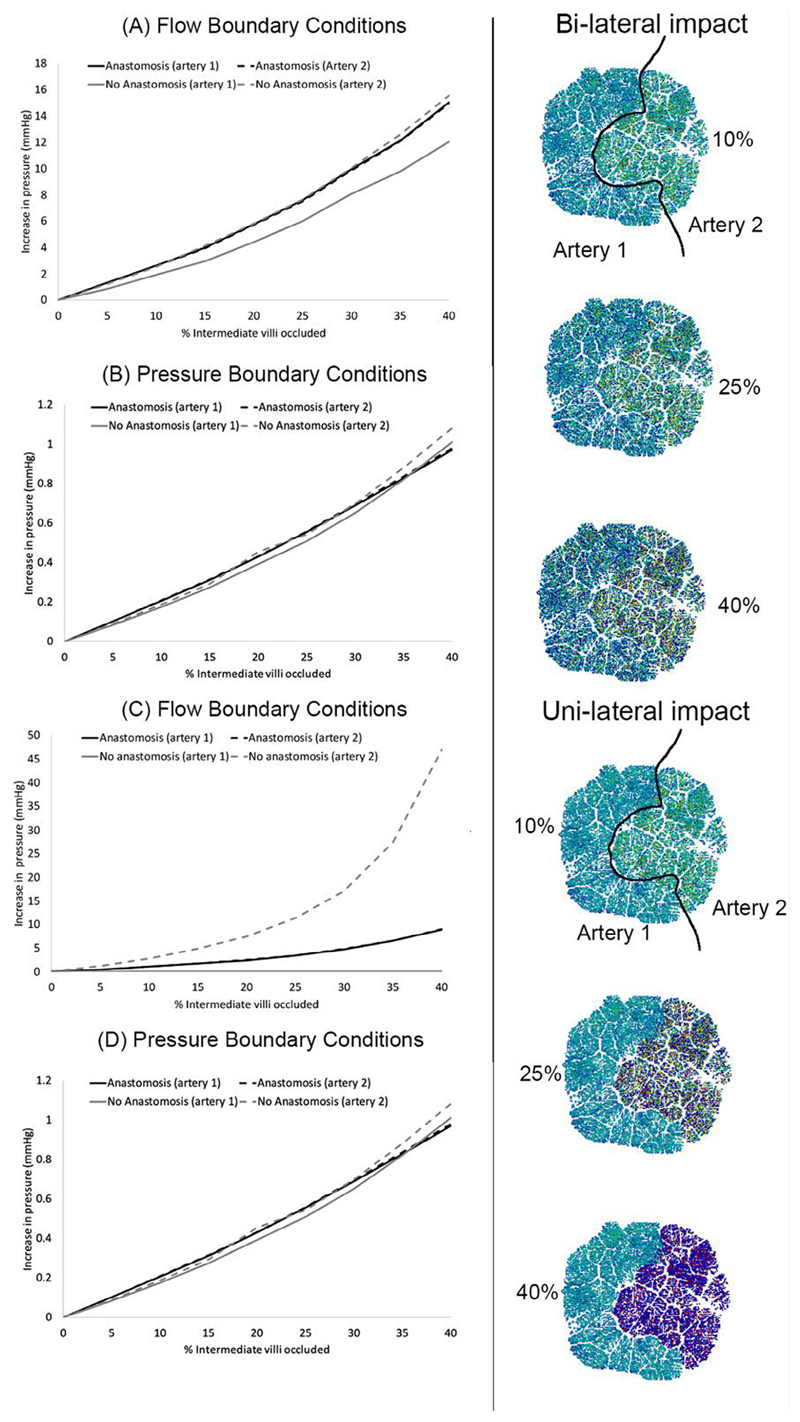
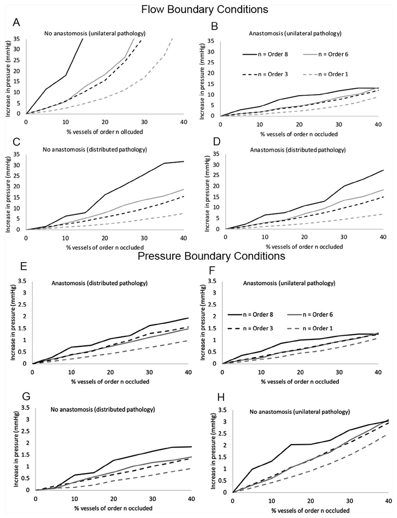
A well-functioning placenta is critical for healthy fetal development, as the placenta brings fetal blood in close contact with nutrient rich maternal blood, enabling exchange of nutrients and waste between mother and fetus. The feto-placental circulation forms a complex branching structure, providing blood to fetal capillaries, which must receive sufficient blood flow to ensure effective exchange, but at a low enough pressure to prevent damage to placental circulatory structures. The branching structure of the feto-placental circulation is known to be altered in complications such as fetal growth restriction, and the presence of regions of vascular dysfunction (such as hypovascularity or thrombosis) are proposed to elevate risk of placental pathology. Here we present a methodology to combine micro-computed tomography and computational model-based analysis of the branching structure of the feto-placental circulation in ex vivo placentae from normal term pregnancies. We analyse how vascular structure relates to function in this key organ of pregnancy; demonstrating that there is a 'resilience' to placental vascular structure-function relationships. We find that placentae with variable chorionic vascular structures, both with and without a Hyrtl's anastomosis between the umbilical arteries, and those with multiple regions of poorly vascularised tissue are able to function with a normal vascular resistance. Our models also predict that by progressively introducing local heterogeneity in placental vascular structure, large increases in feto-placental vascular resistances are induced. This suggests that localised heterogeneities in placental structure could potentially provide an indicator of increased risk of placental dysfunction.

Keywords: Computational model; Haemodynamics; Micro-CT; Placenta.


Conflict of interest statement

Conflict of Interest No benefits in any form have been or will be received from a commercial party related directly or indirectly to the subject of this manuscript.

Figures

Fig. 1
Fig. 1
(A) A schematic of the placental vasculature starting from the umbilical cord - two umbilical arteries in blue carrying deoxygenated blood from the fetus and an umbilical vein in red carrying oxygenated blood from the placenta to the fetus. Vessels branch through the chorionic plate and form stem villi which travel towards the basal plate branching into villous trees with arterioles and venules joined by capillary convolutes at terminal villi. (B) A schematic illustrating Shrahler ordering, the method used to quantify placental branching structure. Terminal vessels are labelled order 1 and by moving from terminal vessels to the inlet(s) order is incremented by one each time two vessels of the same order meet.
Fig. 2
Fig. 2
Maximum intensity projections of A) Placenta 1, and B) Placenta 2. Placentae are shown as anaglyphs from which 3D renderings can be visualised.
Fig. 3
Fig. 3
A summary of methods used to generate a placental vascular structure, illustrated for Placenta 2 (with section numbers in which the methodology is described given in the illustration). (A) Micro-CT images are collected (Pratt et al., 2017) and (B) manually corrected (Aughwane et al., 2019). Then, (C) vascular structures are extracted from imaging via thresholding of pixel intensities. (D) Those vessels that can be identified as distinct connected branches are converted to a graphical network structure, defined by segment centreline location in 3D space and vessel radius. (E) Beyond the level of distinct vascular branches, vascular density was defined based on relative brightness of voxels, and (F) a tree structure was generated using the methods of Clark et al. (Clark et al., 2015). Finally, (G) blood flow and pressure are simulated in the vessel network using a Poiseuille approximation.
Fig. 4
Fig. 4
Micro-CT derived maps of vascularity (voxels identified as belonging to arterial centerlines) for (A) Placenta 1 and (B) Placenta 2 highlights that there is more variability in vascular density locally for Placenta 2, although variability exists in both. This results in a distinct regional structure to branching in a generated vascular tree compared with a tree assuming a homogeneous vascular density. (C, D) are generated trees for Placenta 2 with (C) having homogeneous vascular density, and (D) having heterogeneous vascular density.
Fig. 5
Fig. 5
Model predictions for flow distribution in Placenta 1 and Placenta 2 with flow boundary conditions (equal flow in each umbilical artery) with anastomosis and without an anastomosis present between the umbilical arteries. Colour scale shows volumetric flow in mm3/s.
Fig. 6
Fig. 6
The impact of progressively occluding increasing percentages of order 1 vessels (equivalent to intermediate villi), on blood pressures feeding the placenta in the umbilical arteries. Panels (A, B) show the impact of occlusion in a bilateral manner, and panels (C, D) in a unilateral manner. When an anastomosis is present, the two black lines representing the two umbilical arteries are indistinguishable from one another, however, when the anastomosis is absent there is a pressure differential between the two umbilical arteries and the grey lines representing the two umbilical arteries diverge – this is most pronounced with flow boundary conditions applied to the model (panels A, C) compared with pressure boundary conditions (panels B, D). The right-hand illustrations show diffuse distribution of occluded arteries (bi-lateral impact) in the top three illustrations, and the rbottom three illustrations show the same total percentage occlusion but limited to tissue fed by ‘artery 2’ (uni-lateral impact). Coloured images in the centre show tissue fed by occluded arteries in dark blue.
Fig. 7
Fig. 7
Model predictions of peak umbilical artery blood pressure feeding the placenta for increasing sizes of artery occluded. Results are shown for flow boundary conditions (A-D) and pressure boundary conditions (E-H) The definition of peak umbilical artery pressure is the maximum pressure at the umbilical insertion to the placenta over the two umbilical arteries. Results are shown for (A,E) the case of no anastomosis and unilateral pathology, (B,F) the case of an anastomosis and unilateral pathology, (C,G) no anastomosis with distributed pathology, and (D,H) an anastomosis and distributed pathology. In all cases peak umbilical artery blood pressure is lower when an anastomosis is present than when it is not for the same arterial occlusions.

References

    1. Aughwane R, Schaaf C, Hutchinson JC, et al. Micro-CT and histological investigation of the spatial pattern of feto-placental vascular density. Placenta. 2019;88:36–43. - PMC - PubMed
    1. Aughwane R, Ingram E, Johnstone ED, Salomon LJ, David AL, Melbourne A. Placental MRI and its application to fetal intervention. Prenat Diagn. 2020;40(1):38–48. - PMC - PubMed
    1. Bappoo N, Kelsey LJ, Parker L, et al. Viscosity and haemodynamics in a late gestation rat feto-placental arterial network. Biomech Model Mechan. 2017;16(4):1361–1372. - PubMed
    1. Burton GJ, Jauniaux E. Pathophysiology of placental-derived fetal growth restriction. Am J Obstet Gynecol. 2018;218(2):S745–S761. - PubMed
    1. Clark A, Lin M, Tawhai M, Saghian R, James J. Multiscale modelling of the feto-placental vasculature. Interface Focus. 2015;5(2):20140078. - PMC - PubMed

Publication types

LinkOut - more resources