. 2021 Apr 30;30(6):430-442.
doi: 10.1093/hmg/ddab050.

Loss of nuclear UBE3A activity is the predominant cause of Angelman syndrome in individuals carrying UBE3A missense mutations

Affiliations

Loss of nuclear UBE3A activity is the predominant cause of Angelman syndrome in individuals carrying UBE3A missense mutations

Stijn N V Bossuyt et al. Hum Mol Genet. .

Abstract

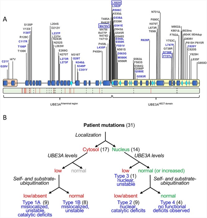
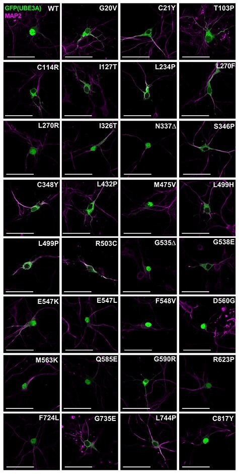
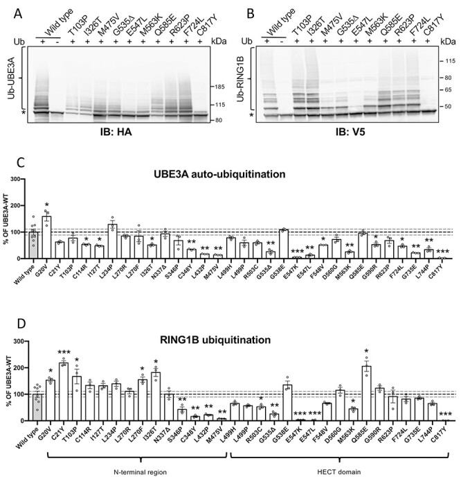
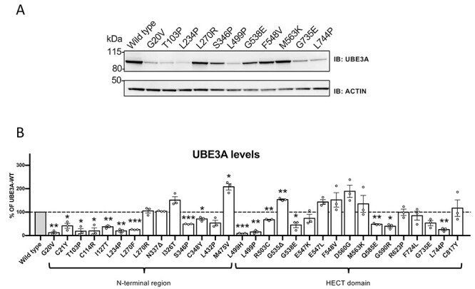
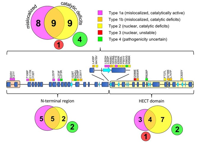
Angelman syndrome (AS) is a severe neurodevelopmental disorder caused by deletion (~75%) or mutation (~10%) of the ubiquitin E3 ligase A (UBE3A) gene, which encodes a HECT type E3 ubiquitin protein ligase. Although the critical substrates of UBE3A are unknown, previous studies have suggested a critical role of nuclear UBE3A in AS pathophysiology. Here, we investigated to what extent UBE3A missense mutations disrupt UBE3A subcellular localization as well as catalytic activity, stability and protein folding. Our functional screen of 31 UBE3A missense mutants revealed that UBE3A mislocalization is the predominant cause of UBE3A dysfunction, accounting for 55% of the UBE3A mutations tested. The second major cause (29%) is a loss of E3-ubiquitin ligase activity, as assessed in an Escherichia coli in vivo ubiquitination assay. Mutations affecting catalytic activity are found not only in the catalytic HECT domain, but also in the N-terminal half of UBE3A, suggesting an important contribution of this N-terminal region to its catalytic potential. Together, our results show that loss of nuclear UBE3A E3 ligase activity is the predominant cause of UBE3A-linked AS. Moreover, our functional analysis screen allows rapid assessment of the pathogenicity of novel UBE3A missense variants which will be of particular importance when treatments for AS become available.


Figures

Figure 1
Figure 1
Functional characterization of UBE3A missense mutations linked to AS. (A) Top: schematic representation of human UBE3A-Isoform 1 (accession no.: NP_570853.1) depicting secondary structure elements, functional domains [amino-terminal Zn-finger of Ube3a ligase (AZUL, orange) and C-terminal HECT domain (yellow)] and the location of AS-linked missense mutations. Missense mutations analyzed in this study are shown in blue and include three novel mutations (boxed). Bottom: schematic representation of mouse UBE3A-Isoform 3 (accession no.: NP_001029134.1). Green color depicts sequence identity with human UBE3A-Isoform 1 (96%). The location of non-conservative and conservative amino acid changes are indicated by red lines and by dashed lines, respectively (see Supplementary Material, Fig. S1 for a detailed sequence alignment). (B) Experimental approach used to assess how missense mutations disrupt UBE3A function. Mutations were introduced in nuclear UBE3A C-terminally tagged with GFP and transfected into mouse primary neurons to determine their subcellular localization. More than half of the mutants appeared to be mislocalized to the cytosol (Type 1). All mutants (in the context of UBE3A without any tag) were also expressed in HEK293T cells to assess their stability. All cytosolic UBE3A mutants appeared unstable while the nuclear localized mutants exhibited normal (or increased) UBE3A levels with the exception of one nuclear mutant that showed low protein levels (Type 3). Lastly, catalytic activity was determined for all mutants. Catalytic deficits were observed in both cytosolic UBE3A mutants (Type1A) and nuclear UBE3A mutants (Type 2). Four mutants did not show any functional deficits in these three assays and were classified Type 4, pathogenicity uncertain.
Figure 2
Figure 2
The majority of tested UBE3A missense mutants mislocalize to the cytosol. Patient-linked missense mutations were introduced at the analogous position in mUBE3A-Iso3 tagged C-terminally with GFP. Neurons were transfected at DIV7 with the indicated UBE3A-GFP constructs and were fixed and stained at DIV10. Neurons were stained for MAP2 (pink) and GFP (UBE3A) was visualized by direct fluorescence (green). Scale bar 50 mm.
Figure 3
Figure 3
AS-associated missense mutations in both the N-terminal region and HECT domain of UBE3A affect catalytic activity. (A and B) Auto-ubiquitination and target ubiquitination of a representative set of UBE3A mutants and quantification of all mutants (C and D). Escherichia coli cells expressing E1, UbcH5 (E2) and with (+) or without (−) ubiquitin were transfected with a plasmid expressing mouse HA-tagged mUBE3A-iso3 (wild-type or the indicated mutant) and V5-RING1B-I53S, a catalytically inactive RING1B mutant that is unable to ubiquitinate itself. Cell lysates were analyzed by SDS-PAGE and immunoblotting using HA antibody to visualize UBE3A (A) and V5 antibody to visualize RING1B (B). Slower migrating, ubiquitinated UBE3A and RING1B species are indicated by a bracket and the unmodified protein band by an asterisk. Quantification of UBE3A auto-ubiquitination (WT, n = 9; mutants, n = 3) (C) and RING1B ubiquitination (WT, n = 8; mutants, n = 3) (D). Values are shown as the mean percent ± SEM of WT UBE3A. Unpaired Student’s t-test (two-tailed). Significant effects are indicated as *P < 0.05, **P < 0.005 and ***P < 0.001. Dashed grey lines depict 95% confidence interval. See Material and Methods section for detailed quantification and statistical methods.
Figure 4
Figure 4
Protein levels of UBE3A missense mutants in HEK293T cells. (A) Protein levels of a representative set of UBE3A mutants and quantification of all mutants (B). HEK293T cells were transiently transfected with untagged UBE3A constructs and protein levels were visualized by anti-UBE3A western blotting using actin as a loading control (A). Quantification of protein levels of all UBE3A variants (n = 3) (B). All data represent mean ± SEM and are normalized against actin. Wild-type (WT) UBE3A levels were set to 100%. One-sample Student’s t-test. Significant effects are indicated as *P < 0.05, **P < 0.005 and ***P < 0.001. See Material and Methods section for detailed quantification and statistical methods.
Figure 5
Figure 5
Classification and distribution of AS-linked UBE3A missense mutations. Schematic representation of human UBE3A-Isoform 1 depicting the location of the 31 AS-linked missense mutations analyzed in this study. Each mutation is color-coded indicating the mechanism by which a mutation disrupts UBE3A function:(i) mislocalization of UBE3A with normal catalytic activity (Type 1A, magenta) or reduced catalytic activity (Type 1B, orange), (ii) reduced catalytic activity of nuclear localized UBE3A (Type 2, yellow) or (iii) reduced stability of nuclear localized UBE3A (Type 3, red). UBE3A mutants that showed no functional deficits in our assays are classified as ‘pathogenicity uncertain’ (Type 4, green). The number of mutants in each class is indicated within the colored circles for the full length UBE3A protein (top) and for the UBE3A N-terminal region and HECT domain separately (bottom).

References

    1. Buiting, K., Williams, C. and Horsthemke, B. (2016) Angelman syndrome — insights into a rare neurogenetic disorder. Nat. Rev. Neurol., 12, 584–593. - PubMed
    1. Matsuura, T., Sutcliffe, J.S., Fang, P., Galjaard, R.J., Jiang, Y.-H., Benton, C.S., Rommens, J.M. and Beaudet, A.L. (1997) De novo truncating mutations in E6-AP ubiquitin-protein ligase gene (UBE3A) in Angelman syndrome. Nat. Genet., 15, 74–77. - PubMed
    1. Kishino, T., Lalande, M. and Wagstaff, J. (1997) UBE3A/E6-AP mutations cause Angelman syndrome. Nat. Genet., 15, 70–73. - PubMed
    1. Scheffner, M. and Kumar, S. (2014) Mammalian HECT ubiquitin-protein ligases: biological and pathophysiological aspects. Biochim. Biophys. Acta, 1843, 61–74. - PubMed
    1. Dindot, S.V., Antalffy, B.A., Bhattacharjee, M.B. and Beaudet, A.L. (2008) The Angelman syndrome ubiquitin ligase localizes to the synapse and nucleus, and maternal deficiency results in abnormal dendritic spine morphology. Hum. Mol. Genet., 17, 111–118. - PubMed

Publication types

Substances