. 2021 Feb 19;12(1):1190.
doi: 10.1038/s41467-021-21464-1.

Mitochondrial DNA editing in mice with DddA-TALE fusion deaminases

Affiliations

Mitochondrial DNA editing in mice with DddA-TALE fusion deaminases

Hyunji Lee et al. Nat Commun. .

Abstract

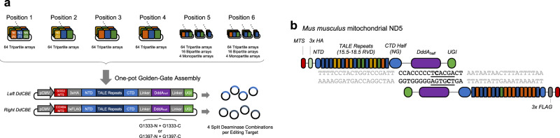
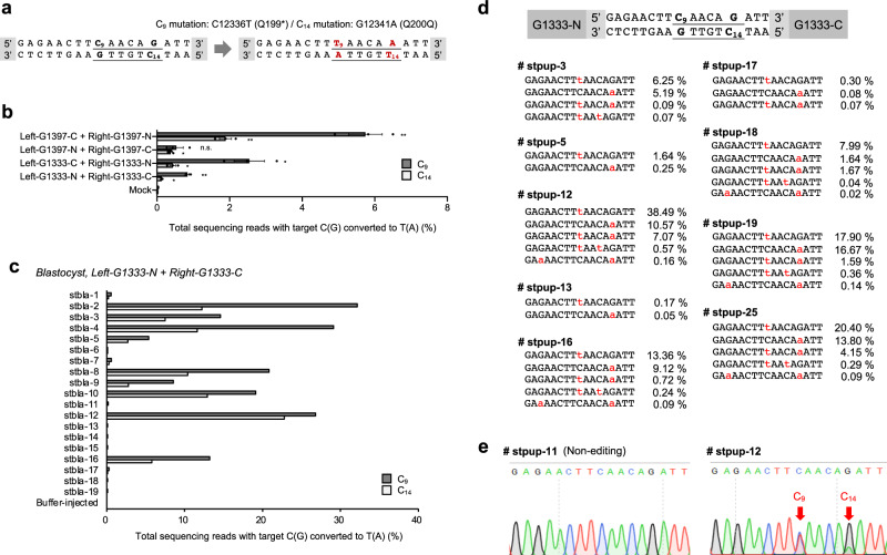
DddA-derived cytosine base editors (DdCBEs), composed of the split interbacterial toxin DddAtox, transcription activator-like effector (TALE), and uracil glycosylase inhibitor (UGI), enable targeted C-to-T base conversions in mitochondrial DNA (mtDNA). Here, we demonstrate highly efficient mtDNA editing in mouse embryos using custom-designed DdCBEs. We target the mitochondrial gene, MT-ND5 (ND5), which encodes a subunit of NADH dehydrogenase that catalyzes NADH dehydration and electron transfer to ubiquinone, to obtain several mtDNA mutations, including m.G12918A associated with human mitochondrial diseases and m.C12336T that incorporates a premature stop codon, creating mitochondrial disease models in mice and demonstrating a potential for the treatment of mitochondrial disorders.


Conflict of interest statement

J.-S.K. is a cofounder of and holds stock in ToolGen. The other authors declare no competing interests.

Figures

Fig. 1
Fig. 1. Schematic illustration for assembling DdCBE and its mitochondrial DNA editing.
a Scheme of one-pot Golden-Gate assembly for efficient DdCBE construction. A total of 424 arrays (64 tripartite arrays × 6 + 16 bipartite arrays × 2 + 4 monopartite arrays × 2) and expression vector were mixed to generate left and right modules for the final plasmid constructs. b Illustration of DdCBE interacting with mouse mitochondrial DNA target ND5. TALE-binding regions are shown in gray and base editing windows are depicted in black. Different repeat variable diresidue modules are shown in orange, blue, green, and yellow, which represent “NI” for adenine, “NG” for thymine, “NN” for guanine, and “HD” for cytosine recognition, respectively.
Fig. 2
Fig. 2. Mouse mitochondrial ND5 point mutation generated by DdCBE-derived base editing.
a DdCBE deaminase-mediated cytosine-to-thymine base editing target and efficiency in NIH3T3 cells. In the target sequence, translation codons are underlined and possible editing loci are shown in red. Transfected combinations of DdCBE are annotated as left or right, -G1333 or -G1397, and -N or -C. P values of left-G1333-N + right-G1333-C, left-G1333-C + right-G1333-N, left-G1397-N + right-G1397-C, and left-G1397-C + right-G1397-N for C10 mutation are 0.0012, 0.0003, 0.0014, and 0.0009, and for C13 mutation are 0.0116, 0.0076, 0.0030, and 0.0003, respectively (*p < 0.05 and **p < 0.01 using Student’s two-tailed t test). b Corresponding base editing efficiency in mouse blastocysts. The sequencing data were obtained from blastocysts that developed after zygotes were microinjected with mRNA encoding the left-G1397-N and right-G1397-C DdCBE. c Alignments of mutant sequences from newborn pups. Targeted deep sequencing was performed using genomic DNA isolated from the tail of the newborns immediately after birth, and that from the toe 7 and 14 days after birth. Edited bases are shown in red. Editing frequencies in the mutant mitochondrial genome are shown. d Editing efficiencies in various tissues of an adult F0 mouse (sipup-1). The sequencing data were obtained from each tissue 50 days after birth. In all graphs, the dark and light gray bars represent the frequency of m.C12539T (C10) and m.G12542A (C13) mutations, respectively. Error bars are the standard error of the mean (s.e.m.) for n = 3 biologically independent samples. Source data are provided in the Source data file.
Fig. 3
Fig. 3. Germline transmission of mutant mtDNA.
a To observe the germline transmission of mtDNA mutations, the female F0 (sipup-3) mouse was crossed with a wild-type C57BL6/J male to obtain F1 pups (101, 102), after which targeted deep sequencing was performed. Edited bases are shown in red. Editing frequencies in the mutant mitochondrial genome are shown. b Base editing efficiencies in various tissues from an F1 newborn pup (101), obtained using targeted deep sequencing of genomic DNA. Dark and light gray bars represent the frequency of m.C12539T (C10) and m.G12542A (C13) mutations, respectively. Error bars are the standard error of the mean (s.e.m.) for n = 3 biologically independent samples. Source data are provided in the Source data file.
Fig. 4
Fig. 4. Mouse mitochondrial ND5 G12918A mutation induced by DdCBE.
a The DdCBE target for generating the m.G12918A point mutation, which would create a D393N change in the ND5 protein. The target codon is underlined and the possible editing locus is shown in red. b The efficiency of cytosine-to-thymine base editing with DdCBE in NIH3T3 cells. The annotations indicate the combination of DdCBE pairs that were co-transfected. Error bars are s.e.m. for n = 3 biologically independent samples (n.s. not significant, *p < 0.05, and **p < 0.01 using Student’s two-tailed t test). P values of left-G1333-N + right-G1333-C, left-G1333-C + right-G1333-N, left-G1397-N + right-G1397-C, and left-G1397-C + right-G1397-N for C6 mutation are 0.0052, 0.0099, 0.0027, and 0.0040, respectively. P values for n.s. is 0.4971. c m.G12918A point mutation base editing efficiency in mouse blastocysts. The sequencing data were obtained from cultured blastocysts that developed after one-cell stage embryos were microinjected with mRNA encoding the left-G1397-C and right-G1397-N DdCBE. d Mice (F0) carrying an ND5 point mutation. F0 pups, which harbor an ND5 point mutation, that developed after microinjection of the DdCBE mRNAs. Corresponding alignment of mutant sequences from newborn pups. Edited bases are shown in red, and the column on the right indicates the editing frequencies in the mutant mitochondrial genome. Source data are provided in the Source data file.
Fig. 5
Fig. 5. Mouse mitochondrial ND5 nonsense mutation generated via cytidine-deaminase-mediated base editing.
a The DdCBE target for generating the m.C12336T nonsense mutation and m.G12341A silent mutation. The m.C12336T (C9) mutation creates a Q199stop mutation in the ND5 protein, whereas m.G12341A (C14) causes a silent Q200Q mutation. Translation triplets are underlined and possible editing loci are shown in red. b The efficiency of the cytosine-to-thymine base editing that creates a nonsense mutation in NIH3T3 cells. The annotations indicate the combination of DdCBE pairs that were co-transfected into cells. Dark and light gray bars represent the frequency of m.C12336T (C9) and m.G12341A (C14) mutations, respectively. Error bars represent s.e.m. for n = 3 biologically independent samples (n.s. not significant, *p < 0.05, and **p < 0.01 using Student’s two-tailed t test). P values of left-G1333-N + right-G1333-C, left-G1333-C + right-G1333-N, left-G1397-N + right-G1397-C, and left-G1397-C + right-G1397-N for C9 mutation are 0.0065, 0.1143, 0.0266, and 0.0037, and for C14 mutation are 0.0077, 0.0144, 0.0406, and 0.0214, respectively. c Base editing efficiency in mouse blastocysts. The sequencing data were obtained from blastocysts that developed after zygotes were microinjected with mRNA encoding left-G1333-N and right-G1333-C DdCBE. Dark and light gray bars represent the frequency of the C9 and C14 mutations, respectively. d Alignment of mutant sequences from newborn pups. Edited bases are shown in red, and the column on the right indicates the editing frequencies in the mutant mitochondrial genome. e Sanger sequencing chromatograms from non-edited and edited mice. The red arrows indicates the substituted nucleotides. Source data are provided in the Source data file.

References

    1. Craven L, Alston CL, Taylor RW, Turnbull DM. Recent advances in mitochondrial disease. Annu. Rev. Genomics Hum. Genet. 2017;18:257–275. doi: 10.1146/annurev-genom-091416-035426. - DOI - PubMed
    1. Schon EA, DiMauro S, Hirano M. Human mitochondrial DNA: roles of inherited and somatic mutations. Nat. Rev. Genet. 2012;13:878–890. doi: 10.1038/nrg3275. - DOI - PMC - PubMed
    1. Gorman GS, et al. Mitochondrial diseases. Nat. Rev. Dis. Prim. 2016;2:16080. doi: 10.1038/nrdp.2016.80. - DOI - PubMed
    1. Hashimoto M, et al. MitoTALEN: a general approach to reduce mutant mtDNA loads and restore oxidative phosphorylation function in mitochondrial diseases. Mol. Ther. 2015;23:1592–1599. doi: 10.1038/mt.2015.126. - DOI - PMC - PubMed
    1. Bacman SR, Williams SL, Pinto M, Peralta S, Moraes CT. Specific elimination of mutant mitochondrial genomes in patient-derived cells by mitoTALENs. Nat. Med. 2013;19:1111–1113. doi: 10.1038/nm.3261. - DOI - PMC - PubMed

Publication types