Review
. 2021 Sep;14(5):978-990.
doi: 10.1038/s41385-020-00370-7. Epub 2021 Feb 19.

Cellular and functional heterogeneity of the airway epithelium

Affiliations
Review

Cellular and functional heterogeneity of the airway epithelium

Jordan D Davis et al. Mucosal Immunol. 2021 Sep.

Erratum in

Abstract

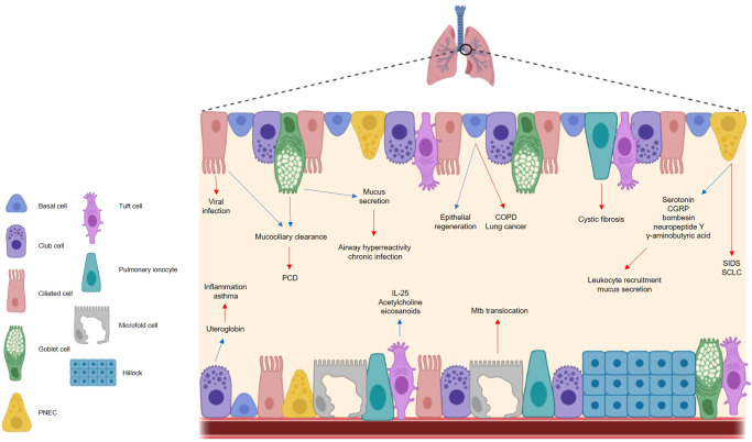
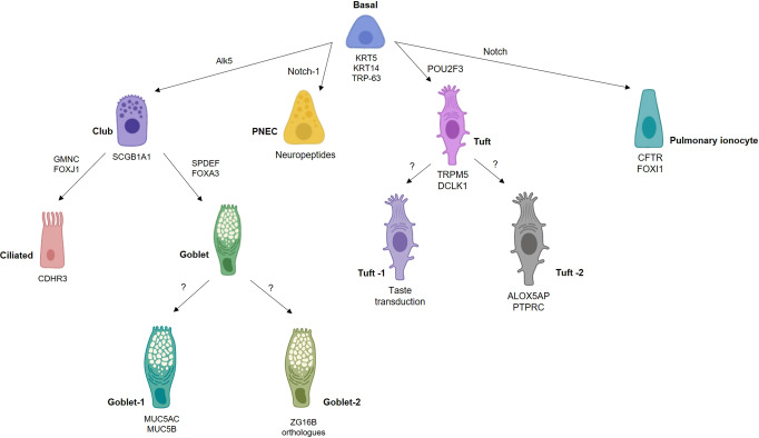
The airway epithelium protects us from environmental insults, which we encounter with every breath. Not only does it passively filter large particles, it also senses potential danger and alerts other cells, including immune and nervous cells. Together, these tissues orchestrate the most appropriate response, balancing the need to eliminate the danger with the risk of damage to the host. Each cell subset within the airway epithelium plays its part, and when impaired, may contribute to the development of respiratory disease. Here we highlight recent advances regarding the cellular and functional heterogeneity along the airway epithelium and discuss how we can use this knowledge to design more effective, targeted therapeutics.


Conflict of interest statement

The authors declare no competing interests.

Figures

Fig. 1
Fig. 1. The respiratory epithelium is important in maintaining respiratory homeostasis but can become implicated in disease.
The cells of the airway epithelium each play a functionally distinct role in health and disease. In a healthy state (blue arrows), basal cells are the principal stem cells of the airway, facilitating epithelial regeneration. Club cells secrete the anti-inflammatory protein uteroglobin, and ciliated cells ensure effective mucociliary clearance in conjunction with goblet cells, the chief mucus producing cells of the airways. Pulmonary neuroendocrine cells secrete a range of neuropeptides, while tuft cells are thought to secrete IL-25, acetylcholine and eicosanoids, although the exact role of these molecules in a respiratory context is unclear. In a diseased state (red arrows), cells of the respiratory epithelium contribute to different illnesses. Basal cells have been linked to COPD and lung cancer, while uteroglobin deficiencies are seen in asthma sufferers. Ciliated cells are the target of viral infection and impaired cilia functionality can cause issues with mucociliary clearance e.g. PCD. Aberrations in mucus production can cause respiratory complications including chronic infection. Neuropeptides induce mucus secretion and leukocyte recruitment and contribute to the pathogenesis of SIDS and SCLC, and microfold cells facilitate Mtb translocation. The mechanisms by which pulmonary ionocytes contribute to cystic fibrosis are largely unknown, and the impact of tuft cells on respiratory disease are poorly characterized. PNEC pulmonary neuroendocrine cell, PCD primary ciliary dyskinesias, CGRP calcitonin gene-related peptide; SIDS sudden infant death syndrome, SCLC small cell lung carcinoma, Mtb mycobacterium tuberculosis, COPD chronic obstructive pulmonary disease.
Fig. 2
Fig. 2. Different transcriptional networks govern airway epithelial cell fate.
Basal cells are the principal stem cells of the airways, differentiating into most other epithelial cell types. Different cell subsets can be characterized by expression of particular genes, for example, pulmonary ionocytes can be classified as expressing FOXI1 and CFTR, the gene mutated in cystic fibrosis. The transcriptional programs governing certain cell fates remains unclear. ZG16B, zymogen granule protein 16.
Fig. 3
Fig. 3. Pulmonary neuroendocrine cells (PNECs) secrete neuropeptides that have both neurological and immunological effects.
PNECs secrete bombesin/bombesin-related peptide, serotonin, GABA and CGRP, which in a neurological context can cause bronchoconstriction, neuroinhibition and vasodilation, respectively. These same effector molecules also stimulate immune responses. For example, bombesin recruits mast cells, serotonin influences cytokine secretion and leukocyte activity, GABA causes goblet cell hyperplasia, and CGRP elicits mucus secretion and ILC2 activation. CGRP calcitonin gene-related peptide, GABA gamma-aminobutyric acid, ILC2 innate lymphoid cell 2.
Fig. 4
Fig. 4. The airway epithelium responds to viral, bacterial, and allergic attack.
a Virus-associated molecular patterns are recognized by a variety of intracellular pattern recognition receptors including toll-like receptors and rig-like helicases e.g. MDA5 and RIG-I, which stimulates release of type 1 and 3 interferons. Collectively, interferons mediate antiviral immunity by inhibiting viral replication, recruiting immune cells (e.g., T cells, B cells, natural killer cells), and upregulating MHC class I expression, leading to increased CD8+ T cell activity. Activated airway epithelial cells also secrete proinflammatory cytokines IL-1α, IL-1β, and TNF-α. b Bacteria can be recognized by multiple TLRs stimulating the release of antimicrobial peptides including human cathelicidin LL-37, defensins, lactoferrin, lysozyme and SLPI. LL-37 mediates recruitment of monocytes, neutrophils, and CD4+ T cells, while defensins, lactoferrin, and lysozyme can destroy bacteria through different mechanisms. AECs also secrete SLPI, which acts in an autocrine/paracrine manner to protect the epithelium against serine proteases and proteolytic enzymes. In response to bacterial infection airway epithelial cells also release a plethora of chemokines, for example, the neutrophilic chemoattractant CXCL8, as well as CCL2, CCL3, CC4, and CCL5, which mediate recruitment of several leukocytes (e.g., monocytes/macrophages, T cells, DCs, neutrophils and natural killer cells). In response to viral and bacterial co-infection, the respiratory epithelium produces IL-17C, which is thought to stimulate neutrophil activity. c Allergens including HDM and fungi activate AEC-bound TLR-4, eliciting secretion of IL-25, IL-33, and TSLP which together drive the downstream allergic response by inducing TH2 cytokine secretion, mucus production, eosinophilia, and DC and ILC2 activation. AECs also secrete CCL20, a potent dendritic cell chemoattractant, as well as uric acid and ATP. TLR toll-like receptor, MDA5 melanoma differentiation-associated protein 5, RIG-I retinoic acid-inducible gene I, IFN interferon, AECs airway epithelial cells, SLPI secretory leukocyte protease inhibitor, IL interleukin, TSLP thymic stromal lymphopoietin, DC dendritic cell, HDM house dust mite, IgE immunoglobulin E, TNF-α tumor necrosis factor-alpha.

References

    1. Karra N, Swindle EJ, Morgan H. Drug delivery for traditional and emerging airway models. Organs-on-a-Chip. 2019;1:100002. doi: 10.1016/j.ooc.2020.100002. - DOI
    1. Basil MC, et al. The cellular and physiological basis for lung repair and regeneration: past, present, and future. Cell Stem Cell. 2020;26:482–502. doi: 10.1016/j.stem.2020.03.009. - DOI - PMC - PubMed
    1. Montoro DT, et al. A revised airway epithelial hierarchy includes CFTR-expressing ionocytes. Nature. 2018;560:319–324. doi: 10.1038/s41586-018-0393-7. - DOI - PMC - PubMed
    1. Plasschaert LW, et al. A single-cell atlas of the airway epithelium reveals the CFTR-rich pulmonary ionocyte. Nature. 2018;560:377–381. doi: 10.1038/s41586-018-0394-6. - DOI - PMC - PubMed
    1. Vieira Braga FA, et al. A cellular census of human lungs identifies novel cell states in health and in asthma. Nat. Med. 2019;25:1153–1163. doi: 10.1038/s41591-019-0468-5. - DOI - PubMed

Publication types

LinkOut - more resources