. 2021 May 7;127(6):765-774.
doi: 10.1093/aob/mcab017.

A water drop-shaped slingshot in plants: geometry and mechanics in the explosive seed dispersal of Orixa japonica (Rutaceae)

Affiliations

A water drop-shaped slingshot in plants: geometry and mechanics in the explosive seed dispersal of Orixa japonica (Rutaceae)

Lan-Jie Huang et al. Ann Bot. .

Abstract

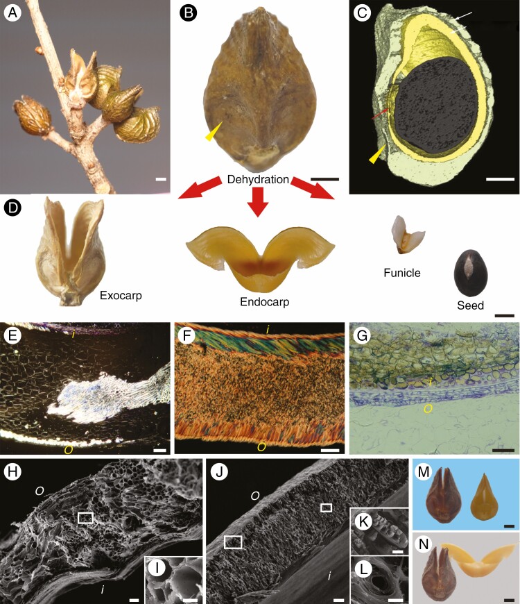
Background and aims: In angiosperms, many species disperse their seeds autonomously by rapid movement of the pericarp. The fruits of these species often have long rod- or long plate-shaped pericarps, which are suitable for ejecting seeds during fruit dehiscence by bending or coiling. However, here we show that fruit with a completely different shape can also rely on pericarp movement to disperse seeds explosively, as in Orixa japonica.

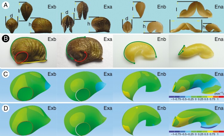
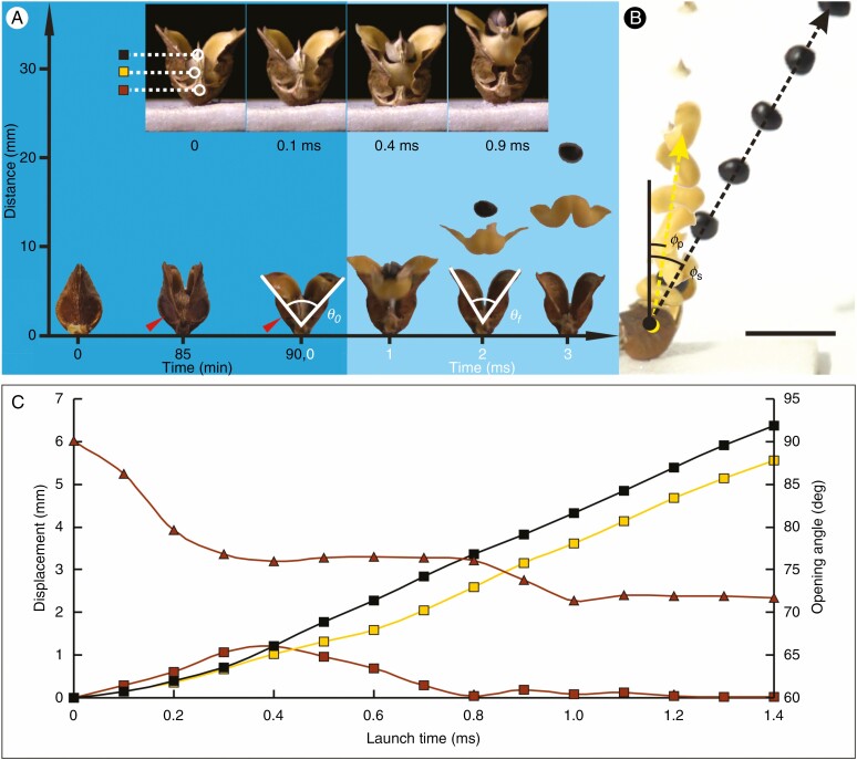
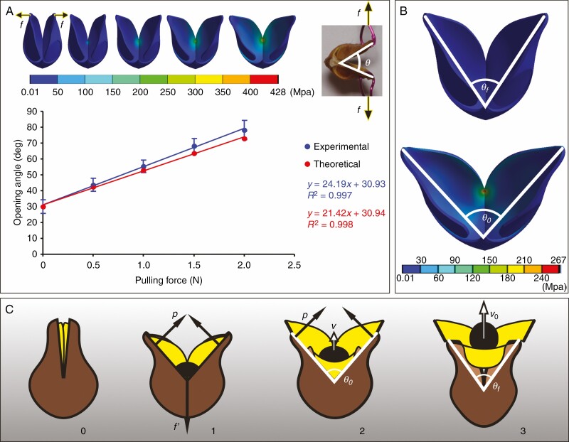
Methods: Fruit morphology was observed by hard tissue sectioning, scanning electron microscopy and micro-computed tomography, and the seed dispersal process was analysed using a high-speed camera. Comparisons were made of the geometric characteristics of pericarps before and after fruit dehiscence, and the mechanical process of pericarp movement was simulated with the aid of the finite element model.

Key results: During fruit dehydration, the water drop-shaped endocarp of O. japonica with sandwich structure produced two-way bending deformation and cracking, and its width increased more than three-fold before opening. Meanwhile the same shaped exocarp with uniform structure could only produce small passive deformation under relatively large external forces. The endocarp forced the exocarp to open by hygroscopic movement before seed launching, and the exocarp provided the acceleration for seed launching through a reaction force.

Conclusions: Two layers of water drop-shaped pericarp in O. japonica form a structure similar to a slingshot, which launches the seed at high speed during fruit dehiscence. The results suggest that plants with explosive seed dispersal appear to have a wide variety of fruit morphology, and through a combination of different external shapes and internal structures, they are able to move rapidly using many sophisticated mechanisms.

Keywords: Orixa japonica; Explosive seed dispersal; Rutaceae; geometry; mechanical simulation; multi-layered structure; plant movement.


Figures

Fig. 1.
Fig. 1.
Fruit morphology of Orixa japonica. (A) Mature aggregate of fruits on a wild plant. (B) Lateral view of a fruitlet on the mature fruit. (C) Three-dimensional reconstruction of the fruitlet showing the internal structure obtained by micro-computed tomography. Yellow arrowheads indicate the basal flat area on the pericarp, in which the exocarp becomes very thin. (D) Parts of a fruitlet after seed dispersal. (E) Polarized light micrograph of a transverse section of the exocarp. Bright vascular tissue is scattered in the dark parenchyma. (F) Polarized light micrograph of a transverse section of the endocarp. The endocarp has a sandwich structure composed of two surface layers of nearly parallel cells and multi-layer thick-walled cells in the middle. Different colours in the two surface layers are interference colours caused by different arrangement directions of cell walls. (G) Light micrograph of a transverse section of the testa (brown) and membranous funiculus (violet) stained with 0.5% toluidine blue. (H) Scanning electron micrograph of a cross-section of the exocarp. (I) Magnification of the boxed area in H. (J) Scanning electron micrograph of a cross-section of the endocarp. (K) Magnification of the left boxed area in J. (L) Magnification of the right boxed area in J. (M) Exocarp and endocarp after rehydration. (N) Exocarp and endocarp after being fully dried. o, outside the fruit; i, inside the fruit. Scale bars: A, B, C, D, M and N = 1 mm; E, F, G, H and J = 50 μm; I, K and L = 10 μm.
Fig. 2.
Fig. 2.
Geometric properties of the pericarp of Orixa japonica. (A) Three-view images of the pericarp before and after fruit dehiscence under the stereomicroscope: l, d and h correspond to the length, width and height of the pericarp, respectively. (B) Continuity of pericarp surfaces before and after fruit dehiscence. The pericarp images were obtained using a stereomicroscope. Green, yellow and red curves represent continuous dorsal suture, ventral suture and boundary line of the jointing area, respectively. (C) Gauss curvature distribution of pericarp surfaces obtained by Mimics and Catia v5 software. The lower right ruler indicates the range of curvature values corresponding to different colours on the surface of the pericarp. (D) Mean curvature distribution of pericarp surfaces obtained by Mimics and Catia v5 software. Ena, dry endocarp after fruit dehiscence; Enb, wet endocarp before fruit dehiscence; Exa, dry exocarp after fruit dehiscence; Exb, wet exocarp before fruit dehiscence.
Fig. 3.
Fig. 3.
Seed launching process of Orixa japonica. (A) Frontal view of the seed launching process. The horizontal digital axis indicates the time of each state in the seed launching process. The zero time in the dark blue area is when the pericarp begins to crack, and the zero time in the light blue area is when the membranous funiculus in the basal joining area is disconnected from the exocarp. The vertical digital axis indicates the height from the base of the pericarp, θ0 is the critical angle of exocarp opening at the beginning of seed launch, and θf is the opening angle of the exocarp after the seed has left the fruitlet. The red arrow indicates the basal fracture area of the exocarp. The four small inset images show details of endocarp detachment from the exocarp during the first millisecond. The four images show in turn the states of the fruit when the seed is about to be launched, when the funiculus is separated from the exocarp, when the seed is separated from the endocarp, and when the upper arms of the endocarp are separated from the exocarp. In the first small image, three coloured squares indicate different parts of the fruit (black, centre of seed; yellow, ventral base of endocarp; brown, ventral base of exocarp), which are used in C. (B) Composite image of the fruit as the seed is launched by the fruit taken from a 1000-fps video in side direction with 1-cm bar. Each image is 1 ms apart. Yellow and black dotted lines represent flight tracks of the endocarp and seed, respectively, φs is the angle between the seed’s flight direction and fruit stalk axis, and φp is the angle between the endocarp flight direction and fruit stalk axis after seed launch. (C) Changes in opening angle of the exocarp and displacements of three different parts of the fruit during seed launching. The triangles represent opening angles of the exocarp during seed launching; the coloured squares represent the displacements of different parts of the fruit indicated in A. Scale bar = 10 mm.
Fig. 4.
Fig. 4.
Biomechanical analysis of pericarp movement during explosive seed dispersal in Orixa japonica. (A) Opening angles of the exocarp under different pulling forces in the tensile test are compared to theoretically predicted values in the finite-element model (FEM) simulation. The red and blue lines are the linear fitting curves of experimental and theoretical data respectively, and the fitting equations and R2 values are displayed on the right-hand side. Error bars of the blue points show the standard deviation of the experimental data. The upper left inset shows the variation of the equivalent stress distribution on the FEM of the exocarp as the tensile load on both sides of the exocarp increases in the simulation, and the upper right insert shows the opening state of the exocarp under pulling force f in the tensile test. (B) Changes of exocarp state and equivalent stress distribution on the exocarp with an opening angle from θf to θ0 in the FEM simulation of exocarp opening. (C) Cartoons of the mechanism of seed launch showing the effect of the interaction between the endocarp (yellow) and the exocarp (brown) on seed (black) acceleration. The numerical sequence below indicates the change of fruit state during seed launching. p, the propulsive force exerted by both sides of the exocarp on the endocarp; f′, the tension exerted by the base of the exocarp on the base of the endocarp before the membranous funiculus breaks from the exocarp; v, the velocity of seed movement; v0, the velocity of seed movement when it leaves the endocarp; θ0, the critical angle of exocarp opening at the beginning of seed launch; θf, the opening angle of the exocarp after seed leaves the fruitlet.

Similar articles

References

    1. Armon S, Efrati E, Kupferman R, Sharon E. 2011. Geometry and mechanics in the opening of chiral seed pods. Science (New York, N.Y.) 333: 1726–1730. - PubMed
    1. Audoly B, Pomeau Y. 2010. Elasticity and geometry: from hair curl to the nonlinear behavior of shells. Oxford: Oxford University Press.
    1. Bar-On B, Sui X, Livanov K, et al. . 2014. Structural origins of morphing in plant tissues. Applied Physics Letters 105: 033703.
    1. Chen PY, McKittrick J, Meyers MA. 2012. Biological materials: Functional adaptations and bioinspired designs. Progress in Materials Science 57: 1492–1704.
    1. Day JS. 2000. Anatomy of capsule dehiscence in sesame varieties. The Journal of Agricultural Science 134: 45–53.

Publication types

LinkOut - more resources