. 2021 May;141(5):681-696.
doi: 10.1007/s00401-021-02263-w. Epub 2021 Feb 20.

Distinct amyloid-β and tau-associated microglia profiles in Alzheimer's disease

Affiliations

Distinct amyloid-β and tau-associated microglia profiles in Alzheimer's disease

Emma Gerrits et al. Acta Neuropathol. 2021 May.

Abstract

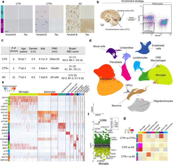
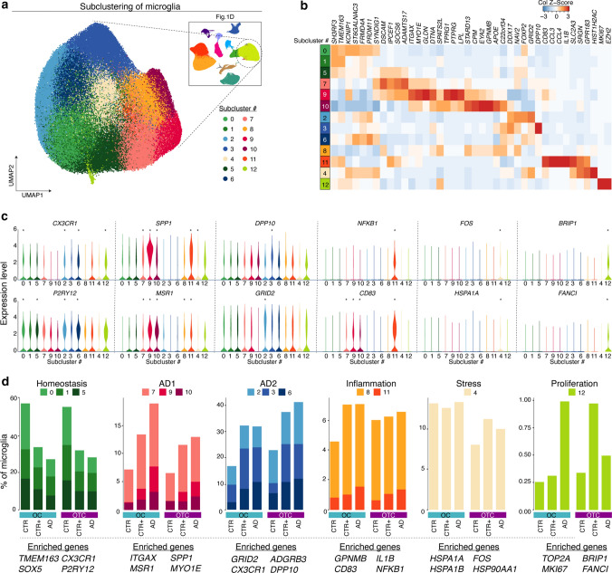
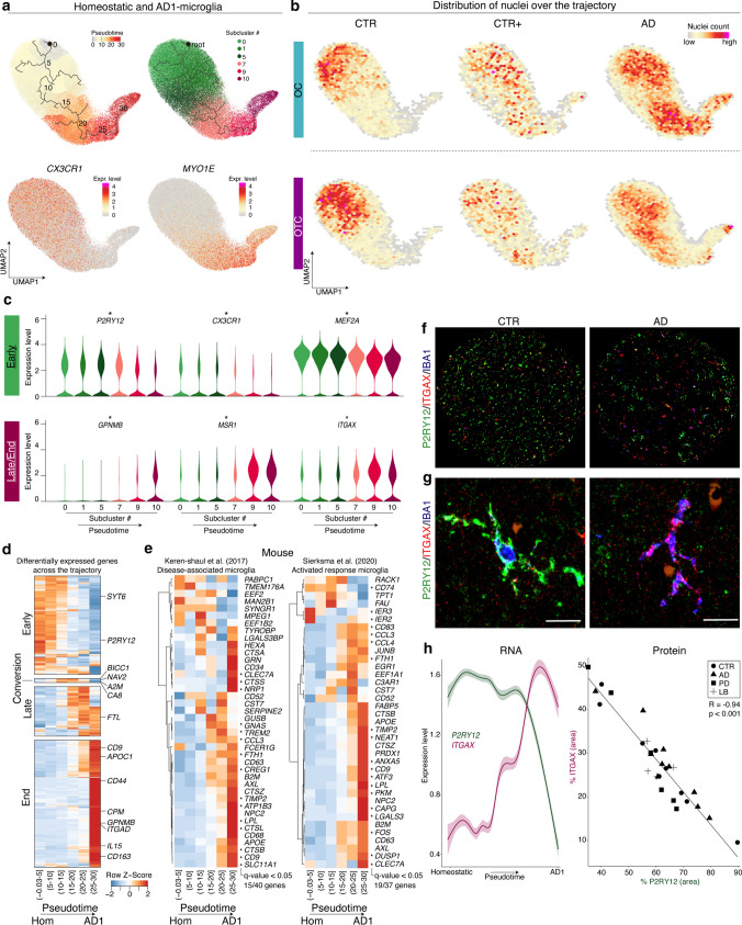
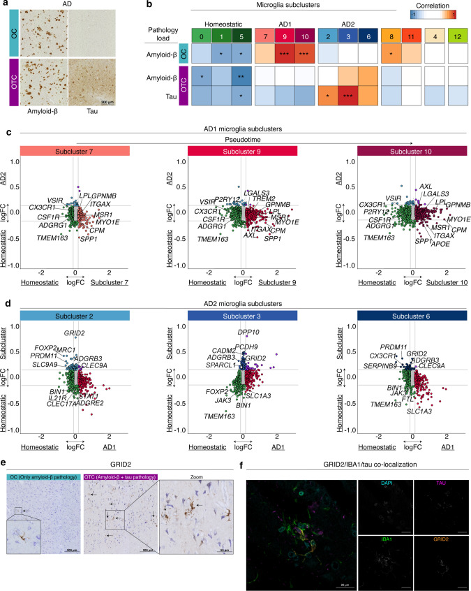
Alzheimer's disease (AD) is the most prevalent form of dementia and is characterized by abnormal extracellular aggregates of amyloid-β and intraneuronal hyperphosphorylated tau tangles and neuropil threads. Microglia, the tissue-resident macrophages of the central nervous system (CNS), are important for CNS homeostasis and implicated in AD pathology. In amyloid mouse models, a phagocytic/activated microglia phenotype has been identified. How increasing levels of amyloid-β and tau pathology affect human microglia transcriptional profiles is unknown. Here, we performed snRNAseq on 482,472 nuclei from non-demented control brains and AD brains containing only amyloid-β plaques or both amyloid-β plaques and tau pathology. Within the microglia population, distinct expression profiles were identified of which two were AD pathology-associated. The phagocytic/activated AD1-microglia population abundance strongly correlated with tissue amyloid-β load and localized to amyloid-β plaques. The AD2-microglia abundance strongly correlated with tissue phospho-tau load and these microglia were more abundant in samples with overt tau pathology. This full characterization of human disease-associated microglia phenotypes provides new insights in the pathophysiological role of microglia in AD and offers new targets for microglia-state-specific therapeutic strategies.

Keywords: Alzheimer’s disease; Amyloid-β; Microglia; Single-nucleus RNA sequencing; Tau.


Conflict of interest statement

MEW, MK, TM and KB are employed by AbbVie, Inc., which has subsidized the study. The other authors declare no competing interests.

Figures

Fig. 1
Fig. 1
Enrichment yields high numbers of microglia and astrocytes for snRNAseq. a Pathological hallmarks of donor groups. b Enrichment strategy for NEUNneg and OLIG2neg nuclei. (Brain Image courtesy of the Neurobiobank of the Institute Born-Bunge, Antwerp (Wilrijk), Belgium (NB190113)). c Donor information. Age, RIN and PMD are presented as mean ± SD. d UMAP depicting 482,472 nuclei derived from 36 human cortical brain samples. Colors indicate cell type clusters. e Heatmap depicting expression of selected cell type marker genes. f Dot plot depicting logFC per gene from the comparison CAM versus microglia nuclei. Size depicts significance level. g Heatmap depicting Chi-squared associations between subcluster distribution within each cell type and donor group per brain region. **: p < 0.01. OC Occipital Cortex; OTC Occipitotemporal Cortex; RIN RNA integrity number; PMD Postmortem delay; CTR non-demented controls; CTR+  non-demented controls with mild amyloid-β pathology; AD clinical and neuropathological Alzheimer’s disease
Fig. 2
Fig. 2
Two groups of microglia subclusters are associated with AD. a UMAP of 148,606 microglia nuclei in 13 subclusters. b Heatmap depicting average expression of three most enriched genes per subcluster. c Violin plots depicting expression of selected genes per subcluster. *: significantly enriched genes for each subcluster compared to all other subclusters (logFC > 0.15, adjusted p-value < 0.05). d Bar plots depicting the percentage of microglia in each subcluster group by category. Representative marker genes are listed on the bottom. OC Occipital Cortex; OTC Occipitotemporal Cortex; CTR non-demented controls; CTR+  non-demented controls with mild amyloid-β pathology; AD clinical and neuropathological Alzheimer’s disease
Fig. 3
Fig. 3
AD1-microglia subclusters gradually transition towards a phagocytic/activated profile. a UMAPs depicting trajectory analysis of homeostatic and AD1-subclusters. Color-scale indicates pseudotime, subclusters and expression of CX3CR1 (homeostasis) and MYO1E (AD1). b Density heatmaps depicting the distribution of nuclei over the UMAP for each sample group. c Violin plots depicting expression of selected genes per subcluster. *: genes significantly differentially expressed (Moran’s I test, q-value < 0.05). d Heatmap depicting all genes significantly differentially expressed over the trajectory *: Moran’s I test, q-value < 0.05). e Heatmaps depicting top 40 (non-ribosomal) DAM genes from [18] and top 37 (non-ribosomal) ARM genes from [30] over pseudotime. *: Moran’s I test, q-value < 0.05. f IBA1, P2RY12, and ITGAX co-expression in tissues from a CTR and an AD donor. Tissue core diameter = 1 mm. g Left: Microglia expressing P2RY12 and IBA1, but not ITGAX. Right: Microglia expressing IBA1 and ITGAX, but not P2RY12. Microglia are from the tissue section and exist next to each other. Scale bar = 20 μm. h Left: Gene expression of P2RY12 (green) and ITGAX (pink) along the trajectory. Right: Correlation between % P2RY12pos area and % ITGAXpos area per sample. OC Occipital Cortex; OTC Occipitotemporal Cortex; CTR non-demented controls; CTR+  non-demented controls with mild amyloid-β pathology; AD clinical and neuropathological Alzheimer’s disease; PD Parkinson’s disease; LB Lewy body dementia
Fig. 4
Fig. 4
Microglia segregate into distinct amyloid-β and tau-associated profiles. a Amyloid-β and phospho-tau immunohistochemistry of an AD donor. b Heatmap depicting Pearson correlations of amyloid-β/tau load versus the percentage of microglia located in each subcluster. *: p ≤ 0,05; **: p ≤ 0.01; ***: p ≤ 0.001. c Four-way plots depicting differential gene expression of the indicated AD1 clusters (on x-axis) versus homeostasis (subclusters 0, 1, 5) and logFC of AD2 (subclusters 2, 3, 6) versus homeostasis (subclusters 0, 1, 5) on the y-axis. d Four-way plots depicting differential gene expression of AD1 (subclusters 7, 9, 10) on the x-axis versus homeostasis (subclusters 0, 1, 5) and logFC of the indicated AD2 clusters versus homeostasis (subclusters 0, 1, 5) on the y-axis. e GRID2 expression (brown) in AD samples with only amyloid-β or both amyloid-β and tau pathology. Cresyl violet was used to detect nuclei. f IBA1 (green), GRID2 (orange) and phospho-tau (magenta) colocalization in human AD brain tissue. OC Occipital Cortex; OTC Occipitotemporal Cortex

Comment in

References

    1. Alsema AM, Jiang Q, Kracht L, Gerrits E, Dubbelaar ML, Miedema A, Brouwer N, Woodbury M, Wachter A, Xi HS, Möller T, Biber KP, Kooistra SM, Boddeke EWG, Eggen BJL. Profiling microglia from AD donors and non-demented elderly in acute human post-mortem cortical tissue. bioRxiv. 2020 doi: 10.1101/2020.03.18.995332. - DOI - PMC - PubMed
    1. Anders S, Pyl PT, Huber W. HTSeq–a Python framework to work with high-throughput sequencing data. Bioinformatics. 2015;31:166–169. doi: 10.1093/bioinformatics/btu638. - DOI - PMC - PubMed
    1. Araki K, Meguro H, Kushiya E, Takayama C, Inoue Y, Mishina M. Selective expression of the glutamate receptor channel δ2 subunit in cerebellar purkinje cells. Biochem Biophys Res Commun. 1993;197:1267–1276. doi: 10.1006/BBRC.1993.2614. - DOI - PubMed
    1. van den Bos H, Spierings DCJ, Taudt AS, Bakker B, Porubský D, Falconer E, Novoa C, Halsema N, Kazemier HG, Hoekstra-Wakker K, Guryev V, den Dunnen WFA, Foijer F, Tatché MC, Boddeke HWGM, Lansdorp PM. Single-cell whole genome sequencing reveals no evidence for common aneuploidy in normal and Alzheimer’s disease neurons. Genome Biol. 2016;17:1–9. doi: 10.1186/s13059-016-0976-2. - DOI - PMC - PubMed
    1. Braak H, Alafuzoff I, Arzberger T, Kretzschmar H, Del Tredici K. Staging of Alzheimer disease-associated neurofibrillary pathology using paraffin sections and immunocytochemistry. Acta Neuropathol. 2006;112:389–404. doi: 10.1007/s00401-006-0127-z. - DOI - PMC - PubMed

Publication types

LinkOut - more resources