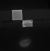
Autofluorescence pattern of parathyroid adenomas
- PMID: 33609395
- PMCID: PMC7893478
- DOI: 10.1093/bjsopen/zraa047
Autofluorescence pattern of parathyroid adenomas
Abstract
Background: Primary hyperparathyroidism (pHPT) is a common endocrine pathology, and it is due to a single parathyroid adenoma in 80-85 per cent of patients. Near-infrared autofluorescence (NIRAF) has recently been used in endocrine surgery to help in the identification of parathyroid tissue, although there is currently no consensus on whether this technique can differentiate between normal and abnormal parathyroid glands. The aim of this study was to describe the autofluorescence pattern of parathyroid adenoma in pHPT.
Methods: Between January and June 2019, patients with pHPT who underwent surgical treatment for parathyroid adenoma were enrolled. Parathyroid autofluorescence was measured.
Results: Twenty-three patients with histologically confirmed parathyroid adenomas were included. Parathyroid adenomas showed a heterogeneous fluorescence pattern, and a well defined autofluorescent 'cap' region was observed in 17 of 23 specimens. This region was on average 28 per cent more fluorescent than the rest of the adenoma, and corresponded to a rim of normal histological parathyroid tissue (sensitivity and specificity 88 and 67 per cent respectively). After resection, all patients were treated successfully, with normal postoperative values of calcium and parathyroid hormone documented.
Conclusion: Parathyroid adenomas show a heterogeneous autofluorescence pattern. Using NIRAF imaging, the majority of specimens showed a well defined autofluorescent portion corresponding to a rim of normal parathyroid tissue. Further studies should be conducted to validate these findings.
Antecedentes: El hiperparatiroidismo primario (primary hyperparathyroidism, pHPT) es una patología endocrina frecuente y su causa es un adenoma paratiroideo único en el 80-85% de los pacientes. La autofluorescencia casi infrarroja (near-infrared auto-fluorescence, NIRAF) ha sido utilizada recientemente en la cirugía endocrina para ayudar en la identificación del tejido paratiroideo, aunque no hay en la actualidad consenso sobre si esta técnica puede diferenciar a las glándulas paratiroideas normales de las anormales. El objetivo de este estudio fue describir el patrón de autofluorescencia del adenoma de paratiroides en el pHPT.
Métodos: Entre enero 2019 y junio 2019, se reclutaron pacientes con pHPT sometidos a tratamiento quirúrgico por adenoma de paratiroides; la autofluorescencia paratiroidea se midió, se describió, y los resultados se analizaron.
Resultados: Se incluyeron 23 pacientes con adenomas de paratiroides confirmados por histología. Los adenomas paratiroideos mostraron un patrón de fluorescencia heterogéneo, y se observó una región capsular bien definida de autofluorescencia en 17 de 23 especímenes. Esta región era por término medio un 28% más fluorescente en comparación con el resto del adenoma y correspondía con el borde de tejido paratiroideo histológicamente normal (sensibilidad y especificidad del 88,2% y 66,7%, respectivamente). Tras la resección, todos los pacientes fueron tratados satisfactoriamente, con valores postoperatorios normales documentados de calcio y PTH.
Conclusiones: Los adenomas de paratiroides muestran un patrón de fluorescencia heterogéneo. Con el uso de NIRAF, la mayoría de los especímenes presentaban una porción de autofluorescencia bien definida correspondiendo al borde de tejido paratiroideo normal. Se deben realizar más estudios para validar estos hallazgos.
© The Author(s) 2021. Published by Oxford University Press on behalf of BJS Society Ltd.
Figures


References
-
- Bilezikian JP, Bandeira L, Khan A, Cusano NE. Hyperparathyroidism. Lancet 2018;391:168–178 - PubMed
-
- Wilhelm SM, Wang TS, Ruan DT, Lee JA, Asa SL, Duh QY et al. The American Association of Endocrine Surgeons guidelines for definitive management of primary hyperparathyroidism. JAMA Surg 2016;151:959–968 - PubMed
-
- Silverberg SJ, Shane E, Jacobs TP, Siris E, Bilezikian JP. A 10-year prospective study of primary hyperparathyroidism with or without parathyroid surgery. N Engl J Med 1999;341:1249–1255 - PubMed
MeSH terms
LinkOut - more resources
Full Text Sources
Other Literature Sources
Miscellaneous

