Review
. 2021 Mar;20(3):222-234.
doi: 10.1016/S1474-4422(20)30440-3.

New insights into atypical Alzheimer's disease in the era of biomarkers

Affiliations
Review

New insights into atypical Alzheimer's disease in the era of biomarkers

Jonathan Graff-Radford et al. Lancet Neurol. 2021 Mar.

Abstract

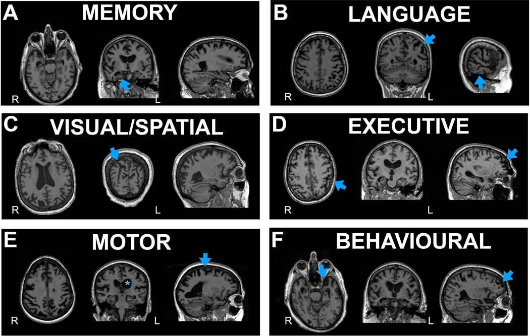
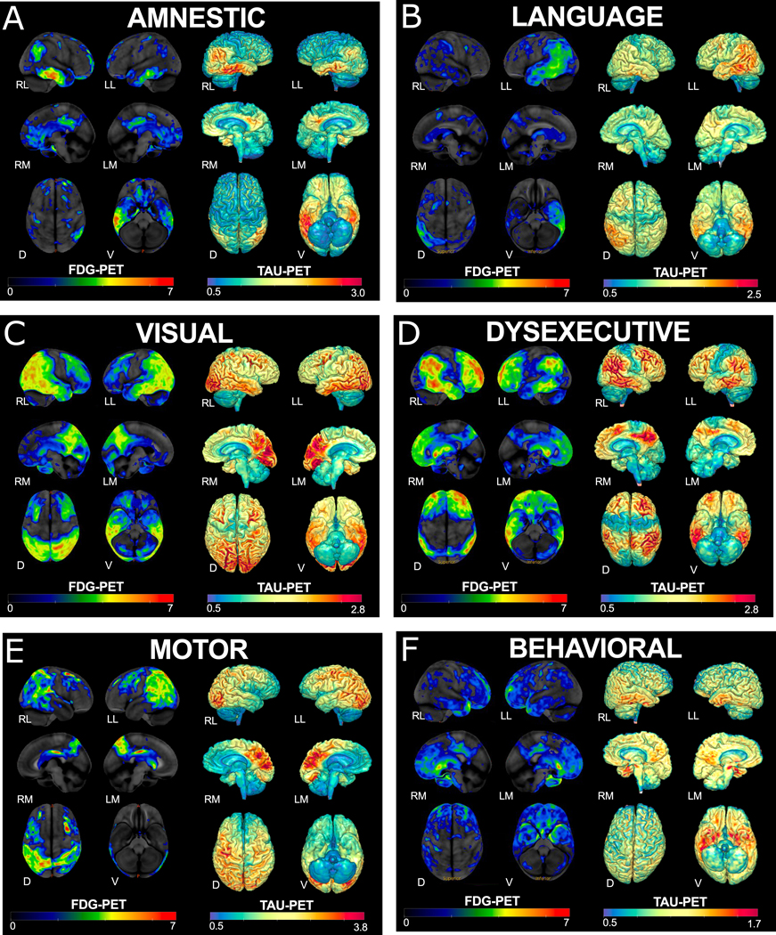
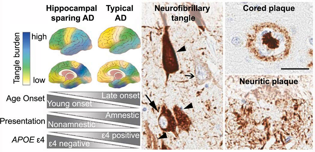
Most patients with Alzheimer's disease present with amnestic problems; however, a substantial proportion, over-represented in young-onset cases, have atypical phenotypes including predominant visual, language, executive, behavioural, or motor dysfunction. In the past, these individuals often received a late diagnosis; however, availability of CSF and PET biomarkers of Alzheimer's disease pathologies and incorporation of atypical forms of Alzheimer's disease into new diagnostic criteria increasingly allows them to be more confidently diagnosed early in their illness. This early diagnosis in turn allows patients to be offered tailored information, appropriate care and support, and individualised treatment plans. These advances will provide improved access to clinical trials, which often exclude atypical phenotypes. Research into atypical Alzheimer's disease has revealed previously unrecognised neuropathological heterogeneity across the Alzheimer's disease spectrum. Neuroimaging, genetic, biomarker, and basic science studies are providing key insights into the factors that might drive selective vulnerability of differing brain networks, with potential mechanistic implications for understanding typical late-onset Alzheimer's disease.


Figures

Figure 1:
Figure 1:. MRI across AD phenotypes
A. Memory (Typical amnestic); blue arrows highlighting hippocampal atrophy B. Language (logopenic variant primary progressive aphasia); blue arrow highlighting left temporal-parietal atrophy C. Visual/Spatial (posterior cortical atrophy); blue arrow indicating parieto-occipital atrophy D. Executive (Dysexecutive); blue arrows indicating frontoparietal atrophy E. Motor (corticobasal syndrome); asterisk highlighting left greater than right hemisphere atrophy and arrow indicating atrophy around the motor cortex F. Behavioural with arrows demonstrating temporal>frontal atrophy
Figure 2:
Figure 2:. FDG and tau PET across AD phenotypes
FDG-PET on left and tau PET on right of representative cases of each AD phenotype: A) Memory (Typical amnestic); B) Language (logopenic variant primary progressive aphasia); C) Visual/Spatial (posterior cortical atrophy); D) Executive (Dysexecutive); E) Motor (corticobasal syndrome); F) Behavioural.The z-scores, relative to a normative database, of pons intensity normalized FDG-PET scans for each individual are displayed on stereotactic surface projections using Cortex ID (GE Healthcare). Red colour indicates greater hypometabolism. The cerebellar crus intensity normalized Tau-PET scan (Tauvid; AV1451; flortaucipir F18; Avid Radiopharmaceuticals, Eli Lilly and Co.) is overlaid on the grey matter segmentations of each subject’s own T1 weighted structural MRI scan. Red colour indicates higher intensity of tracer. RL- Right Lateral, LL – Left Lateral, RM–RightMedial, LM – Left Medial, D – Dorsal, V-Ventral
Figure 3:
Figure 3:
(Left panel) Neuropathologic subtypes of AD are characterized by distribution of neurofibrillary tangle pathology. Illustrations depict the hippocampal sparing subtype with greater cortical pathology relative to sparing of the hippocampus. The typical AD subtype demonstrates expected patterns of both limbic and cortical involvement. Disproportionate differences in age onset, clinical presentation, and APOE ε4 positivity are observed between hippocampal sparing AD and typical AD. (Middle panel) Abnormal accumulation of intracellular tau pathology is observed with increasing severity from pre-tangles (open arrow) to mature tangles (closed arrow). As the neuron dies, a remnant of the tau pathology remains in the extracellular space as ghost tangles (arrowhead). (Right panel) Top: Classic cored plaques are typically observed in AD brains. Bottom: Neuritic plaques can be readily observed using tau antibodies, but may be more easily distinguished by silver stain or thioflavin-S microscopy (not shown).(Scale bar 50μm; the PHF-1 tau antibody was a kind gift from Peter Davies; the 6F/3D amyloid-β antibody was purchased from Dako)

References

    1. Jack CR Jr., Bennett DA, Blennow K, et al. NIA-AA Research Framework: Toward a biological definition of Alzheimer’s disease. Alzheimers Dement. 2018;14:535–62. - PMC - PubMed
    1. Barnes J, Dickerson BC, Frost C, Jiskoot LC, Wolk D, van der Flier WM. Alzheimer’s disease first symptoms are age dependent: Evidence from the NACC dataset. Alzheimers Dement. 2015;11:1349–57. - PMC - PubMed
    1. Balasa M, Gelpi E, Antonell A, et al. Clinical features and APOE genotype of pathologically proven early-onset Alzheimer disease. Neurology. 2011;76:1720–5. - PubMed
    1. Murray ME, Graff-Radford NR, Ross OA, Petersen RC, Duara R, Dickson DW. Neuropathologically defined subtypes of Alzheimer’s disease with distinct clinical characteristics: a retrospective study. Lancet Neurol. 2011;10:785–96. - PMC - PubMed
    1. Winblad B, Amouyel P, Andrieu S, et al. Defeating Alzheimer’s disease and other dementias: a priority for European science and society. Lancet Neurol. 2016;15:455–532. - PubMed

Publication types