. 2021 Jun:155:88-98.
doi: 10.1016/j.yjmcc.2021.02.005. Epub 2021 Feb 18.

Evidence that the acetyltransferase Tip60 induces the DNA damage response and cell-cycle arrest in neonatal cardiomyocytes

Affiliations

Evidence that the acetyltransferase Tip60 induces the DNA damage response and cell-cycle arrest in neonatal cardiomyocytes

Xinrui Wang et al. J Mol Cell Cardiol. 2021 Jun.

Abstract

Tip60, a pan-acetyltransferase encoded by the Kat5 gene, is enriched in the myocardium; however, its function in the heart is unknown. In cancer cells, Tip60 acetylates Atm (Ataxia-telangiectasia mutated), enabling its auto-phosphorylation (pAtm), which activates the DNA damage response (DDR). It was recently reported that activation of pAtm at the time of birth induces the DDR in cardiomyocytes (CMs), resulting in proliferative senescence. We therefore hypothesized that Tip60 initiates this process, and that depletion of Tip60 accordingly diminishes the DDR while extending the duration of CM cell-cycle activation. To test this hypothesis, an experimental model was used wherein a Myh6-driven Cre-recombinase transgene was activated on postnatal day 0 (P0) to recombine floxed Kat5 alleles and induce Tip60 depletion in neonatal CMs, without causing pathogenesis. Depletion of Tip60 resulted in reduced numbers of pAtm-positive CMs during the neonatal period, which correlated with reduced numbers of pH2A.X-positive CMs and decreased expression of genes encoding markers of the DDR as well as inflammation. This was accompanied by decreased expression of the cell-cycle inhibitors Meis1 and p27, activation of the cell-cycle in CMs, reduced CM size, and increased numbers of mononuclear/diploid CMs. Increased expression of fetal markers suggested that Tip60 depletion promotes a fetal-like proliferative state. Finally, infarction of Tip60-depleted hearts at P7 revealed improved cardiac function at P39 accompanied by reduced fibrosis, increased CM cell-cycle activation, and reduced apoptosis in the remote zone. These findings indicate that, among its pleiotropic functions, Tip60 induces the DDR in CMs, contributing to proliferative senescence.

Keywords: Atm; Cell-cycle; DNA damage response; Myocardial infarction; Neonatal cardiomyocytes; Tip60.


Conflict of interest statement

Disclosures

No conflicts of interest, financial or otherwise, are declared by the authors.

Figures
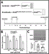
Figure 1.
Figure 1.. Tamoxifen-induced activation of Cre-recombinase at P0 causes Tip60 depletion in the heart at later postnatal stages.
Control (Kat5f/f) and experimental (Kat5f/f;Myh6-merCremer, denoted Kat5Δ/Δ) pups were injected with 250 μg tamoxifen on the day of birth (P0) to initiate recombination of the floxed Kat5 gene. Panel A schematically displays how the control and knockout genotypes are designated in this paper, and the postnatal days when hearts were harvested (H) for analysis. Panel B shows results from qRT-PCR analyses revealing the extent of Kat5 mRNA knockdown on each neonatal day. Panel C is a western blot showing Tip60 protein depletion at P12. Error bars denote ±SEM. Statistical significance was determined using an unpaired two-tailed t-test. *P<0.05 vs Kat5f/f.
Figure 2.
Figure 2.. DDR mitigation in Tip60-depleted neonatal hearts.
Hearts in control (Kat5f/f) and experimental (Kat5Δ/Δ) neonates that were administered tamoxifen at P0 were harvested on the indicated postnatal days. Panel A shows sections that were double-immunostained to detect GATA4 (red) and pAtm (green), with quantitation expressed as percentages of pAtm-positive CMs enumerated by manually scanning entire sections at 1,000x magnification. Panel B shows numbers of pH2A.X CMs in sections double-immunostained to detect pH2A.X and α-actinin. White arrows point to examples of nuclei that are over half-filled with FITC fluorescence and were counted as pH2A.X-positive CMs whereas yellow arrows highlight examples of nuclei that were not counted as pH2A.X-positive. Panel C shows the expression of genes in the DDR pathway (DDR markers), and genes associated with the senescence-associated secretory phenotype (SASP markers), determined using qRT-PCR. Data in panels A-C are presented as mean ±SEM. In panels A-B, numbers (N) of individual hearts evaluated in each group were as follows: P7, 3 vs 4; P12, 7 vs 8; P39, 7 vs 7. Two-way ANOVA revealed that Tip60 depletion was associated with statistically significant reduced levels of pAtm (panel A), pH2A.X (panel B), and the genes denoted by red bars in panel C, across the neonatal timeline. In panel C, symbols * and Ϯ adjacent to individual data points respectively denote P<0.05 vs. Kat5f/f by post-hoc analysis using the Bonferroni multiple comparison test, and by correlation analysis. [Note: This figure requires color reproduction.]
Figure 3.
Figure 3.. Cell-cycle activation in Tip60-depleted neonatal hearts.
Following injection of tamoxifen at P0, hearts harvested on the indicated postnatal days were subjected to the following determinations. Panel A shows results of immunostaining to detect Ki67, BrdU, and pH3, plus GATA4 to identify CMs. Percentages of cell-cycle-activated CMs were enumerated by blinded observers; the entirety of each section was scanned at 1,000x magnification. Panel B shows results of qPCR determinations to detect the expression of genes that activate and inhibit the cell-cycle. Panel C shows assessment of percentages of CMs isolated from P12 hearts that were mononuclear, and mononuclear/diploid (MNDCM). CMs immunostained with cTnT (green) are shown at two magnifications (CM nuclei are pseudo-colored white). Yellow and red arrowheads denote CMs that are mononuclear/diploid and mononuclear/polyploid, respectively. All data are presented as means ±SEM. For Panels A and B, two-way ANOVA revealed that the changes in Ki67, BrdU, pH3, and the genes labeled with red bars (Panel B) in Tip60-depleted hearts across neonatal time-points were statistically significant (P<0.05). P values and asterisks (*P<0.05) next to individual data points report results of post-hoc analysis using a Bonferroni-corrected multiple comparison test. Ϯ next to individual data points denotes P<0.05 vs. Kat5f/f by correlation analysis. For Panel C, data were analyzed by an unpaired, two-tailed Student’s t test. [Note: This figure requires color reproduction.]
Figure 4.
Figure 4.. Increased CM density and expression of fetal gene markers in Tip60-depleted hearts.
Following injection of tamoxifen at P0, hearts were harvested on the indicated postnatal days and subjected to the following determinations. Panel A: WGA staining of Tip60-depleted CMs in transverse sections reveals reduced size and increased density. Panel B: qPCR shows increased expression of fetal gene markers in Tip60-depleted hearts at P39. All data are presented as means ±SEM. Two-way ANOVA revealed that the changes in CM size/density (Panel A) and expression of the genes labeled with red bars (Panel B) in Tip60-depleted hearts across neonatal time-points were statistically significant. Asterisks (*) next to individual data points indicate P<0.05 vs Kat5f/f by post-hoc comparison using a Bonferroni multiple comparison test. Ϯ next to individual data points denotes P<0.05 vs. Kat5f/f by correlation analysis. [Note: This figure requires color reproduction.]
Figure 5.
Figure 5.. Reduced scarring, improved cardiac function, and increased cell-cycle activation after MI in Tip60-depleted hearts.
Kat5f/f and Kat5Δ/Δ hearts treated with tamoxifen on P0 were infarcted on P7 via permanent ligation of the left main coronary artery. On P39, cardiac function was assessed by echocardiography, after which hearts were processed for histology, and transverse sections were removed at equal intervals below the site of ligation. Panel A (upper) shows effects on scarring; quantification of scar size and echocardiographic function are shown below. ‘Control’ denotes hearts that were not infarcted. Panel B shows increased cell-cycle activity in Tip60-depleted CMs; BZ and RZ respectively denote the border and remote zones. In both panels, statistical significance was determined using unpaired, two-tailed Student t tests. Yellow arrowheads denote Ki67/BrdU/pH3-positive CMs. [Note: This figure requires color reproduction.]
Figure 6.
Figure 6.. Increased density, reduced size, and diminished apoptosis of CMs in Tip60-depleted/infarcted hearts at P39.
Kat5f/f and Kat5Δ/Δ hearts were treated with tamoxifen on P0, infarcted on P7, and harvested on P39 followed by histological processing. Panel A: Sections were stained with fluorescently-labeled wheat germ agglutinin (WGA), and areas containing CMs in transverse orientation were photographed and processed with ImageJ to estimate CM size and density within both the infarct border (BZ) and remote zones (RZ). Panel B: Sections were double-stained with fluorescent WGA to identify CM outlines and with anti-caspase-3 to identify apoptotic CMs, followed by enumerating the total number of caspase-positive CMs (examples are denoted by yellow arrowheads) in each section. In both panels, statistical significance was determined using unpaired, two-tailed Student t tests. [Note: This figure requires color reproduction.]

Similar articles

Cited by

References

    1. Sadek H, Olson EN. Toward the Goal of Human Heart Regeneration. Cell Stem Cell. 2020;26(1):7–16. - PMC - PubMed
    1. Galdos FX, Guo Y, Paige SL, VanDusen NJ, Wu SM, Pu WT. Cardiac Regeneration: Lessons From Development. Circ Res. 2017;120(6):941–59. - PMC - PubMed
    1. Fisher JB, Kim MS, Blinka S, Ge ZD, Wan T, Duris C, et al. Stress-induced cell-cycle activation in Tip60 haploinsufficient adult cardiomyocytes. PLoS One. 2012;7(2):e31569. - PMC - PubMed
    1. Hu Y, Fisher JB, Koprowski S, McAllister D, Kim MS, Lough J. Homozygous disruption of the Tip60 gene causes early embryonic lethality. Dev Dyn. 2009;238(11):2912–21. - PMC - PubMed
    1. Numata A, Kwok HS, Zhou QL, Li J, Tirado-Magallanes R, Espinosa Angarcia V, et al. Lysine acetyltransferase Tip60 is required for hematopoietic stem cell maintenance. Blood. 2020. - PMC - PubMed

Publication types

MeSH terms