. 2021 Jan-Jun:296:100432.
doi: 10.1016/j.jbc.2021.100432. Epub 2021 Feb 18.

Structural characterization of a PCP-R didomain from an archaeal nonribosomal peptide synthetase reveals novel interdomain interactions

Affiliations

Structural characterization of a PCP-R didomain from an archaeal nonribosomal peptide synthetase reveals novel interdomain interactions

Sandesh Deshpande et al. J Biol Chem. 2021 Jan-Jun.

Abstract

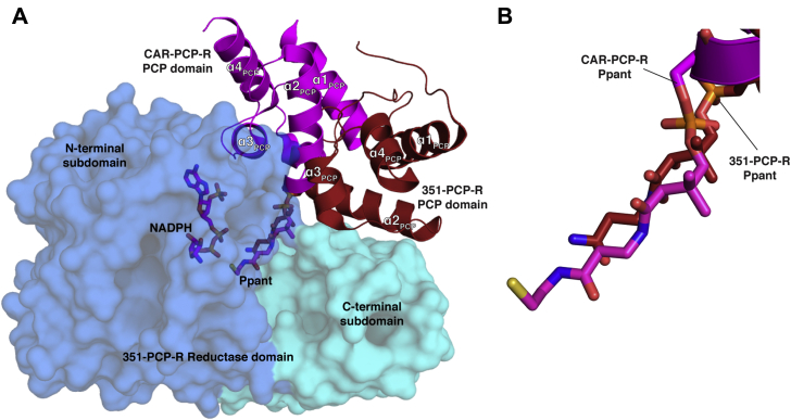
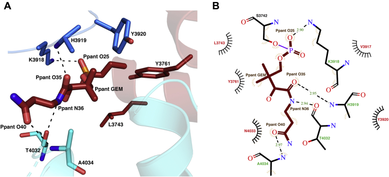
Nonribosomal peptide synthetases (NRPSs) are multimodular enzymes that produce a wide range of bioactive peptides, such as siderophores, toxins, and antibacterial and insecticidal agents. NRPSs are dynamic proteins characterized by extensive interdomain communications as a consequence of their assembly-line mode of synthesis. Hence, crystal structures of multidomain fragments of NRPSs have aided in elucidating crucial interdomain interactions that occur during different steps of the NRPS catalytic cycle. One crucial yet unexplored interaction is that between the reductase (R) domain and the peptide carrier protein (PCP) domain. R domains are members of the short-chain dehydrogenase/reductase family and function as termination domains that catalyze the reductive release of the final peptide product from the terminal PCP domain of the NRPS. Here, we report the crystal structure of an archaeal NRPS PCP-R didomain construct. This is the first NRPS R domain structure to be determined together with the upstream PCP domain and is also the first structure of an archaeal NRPS to be reported. The structure reveals that a novel helix-turn-helix motif, found in NRPS R domains but not in other short-chain dehydrogenase/reductase family members, plays a major role in the interface between the PCP and R domains. The information derived from the described PCP-R interface will aid in gaining further mechanistic insights into the peptide termination reaction catalyzed by the R domain and may have implications in engineering NRPSs to synthesize novel peptide products.

Keywords: X-ray crystallography; archaea; nonribosomal peptide synthetase; peptide biosynthesis; peptide carrier protein domain; protein–protein interaction; reductase domain; short-chain dehydrogenase/reductase; structure–function.


Conflict of interest statement

Conflict of interest The authors declare that they have no conflicts of interest with the contents of this article.

Figures

Figure 1
Figure 1
Ribbon diagram representations of the 351-PCP–R structures.A, the unmodified 351-PCP–R structures. B, the Ppant-modified 351-PCP–R structures (N-terminal reductase subdomain—blue, C-terminal R subdomain—cyan, and PCP domain—red). PCP–R, peptide carrier protein–reductase; Ppant, 4′-phosphopantetheine.
Figure 2
Figure 2
Details of the 351 reductase domain structure.A, features of the 351-PCP–R N-terminal subdomain (N-terminal reductase [R] subdomain—blue, C-terminal R subdomain—cyan, and PCP domain—red). The β-sheet in the cofactor-binding Rossmann fold is shown in magenta, active-site residues are shown as blue sticks, and Ppant is shown as red sticks. B, superimposed N-terminal regions (L3789–N3824 in 351-PCP–R) of various nonribosomal peptide synthetase and carboxylic acid reductase structures (351-PCP–R—blue, AusA-R—orange, carboxylic acid reductase PCP–R—magenta, MxaA-R—yellow, and Mtb-R—green). AusA-R, R domain of a dimodular nonribosomal peptide synthetase (NRPS) from the aureusimine biosynthetic cluster; MxaA-R, R domain of the NRPS module of the myxalamid biosynthetic pathway; PCP–R, peptide carrier protein–reductase; Ppant, 4′-phosphopantetheine; Mtb-R, R domain of a Mycobacterium tuberculosis NRPS.
Figure 3
Figure 3
Details of the 351 reductase domain NADPH-binding site.A, Ppant-modified 351-PCP–R showing patches of positive difference density in the NADPH-binding site as green mesh. The active-site resides are shown in ball and stick representation. NADPH superimposed from structure of MxaA-R (6) is shown. The FoFc map is contoured at 2.5 σ. B, comparison of gating loop orientations (351-PCP–R—blue, MxaA-R—yellow (6), and Mtb-R—green (27)). NADPH from MxaA-R and Ppant from 351-PCP–R are shown in stick representation. MxaA-R, R domain of the nonribosomal peptide synthetase module of the myxalamid biosynthetic pathway; PCP–R, peptide carrier protein–reductase; Ppant, 4′-phosphopantetheine; Mtb-R, R domain of a Mycobacterium tuberculosis nonribosomal peptide synthetase.
Figure 4
Figure 4
The peptide carrier protein–reductase interface is mediated by an interface of helix–turn–helix motif.A, structural alignment of unmodified (gray) and Ppant-modified 351-PCP–R (blue, cyan, and red). The interface–HTH motif is highlighted. B, structural alignment of the interface–HTH motif of all nonribosomal peptide synthetase reductase domain structures (351-PCP–R—cyan, AusA-R—orange (8), MxaA-R—yellow (6), Mtb-R—green (27), and carboxylic acid reductase–PCP–R—magenta (20)). AusA-R, R domain of a dimodular nonribosomal peptide synthetase from the aureusimine biosynthetic cluster; HTH, helix–turn–helix; Mtb-R, R domain of a Mycobacterium tuberculosis nonribosomal peptide synthetase; MxaA-R, R domain of the nonribosomal peptide synthetase module of the myxalamid biosynthetic pathway; PCP–R, peptide carrier protein–reductase; Ppant, 4′-phosphopantetheine.
Figure 5
Figure 5
The Ppant-modified 351-PCP-R structure reveals a previously unobserved conformation.A, the position of PCP domain of 351-PCP–R compared with the PCP domain of CAR–PCP–R (20) following structural alignment of the reductase domains (351-PCP–R—blue, cyan, and red; CAR–PCP–R—magenta). The 351-PCP–R domain is shown in surface representation (blue and cyan). NADPH from CAR–PCP–R (magenta) is shown. B, comparison of the position of Ppant in Ppant-modified 351-PCP–R and CAR–PCP–R following structural alignment of the reductase domains. CAR–PCP–R, carboxylic acid reductase–peptide carrier protein–reductase; Ppant, 4′-phosphopantetheine.
Figure 6
Figure 6
The 351-peptide carrier protein–reductase domain interface. Interacting residues in the peptide carrier protein domain (red), reductase C-terminal subdomain (cyan), and gating loop (blue) are shown in stick representation. Hydrogen bonds are shown as black dotted lines.
Figure 7
Figure 7
The interactions between the Ppant group and 351-peptide carrier protein–reductase protein.A, the interactions between Ppant group and peptide carrier protein domain (red), reductase C-terminal subdomain (cyan), and gating loop residues (blue). Hydrogen bonds are shown as black dotted lines. B, Ppant interface diagram calculated by LigPlot+ (55). Residues involved in hydrogen bonding and hydrophobic interactions with Ppant are represented in green and red, respectively. Hydrogen bonds between atoms are shown as black dotted lines. Ppant, 4′-phosphopantetheine; Ppant GEM, geminal dimethyl group of Ppant.

References

    1. Marahiel M.A., Stachelhaus T., Mootz H.D. Modular peptide synthetases involved in nonribosomal peptide synthesis. Chem. Rev. 1997;97:2651–2673. - PubMed
    1. Süssmuth R.D., Mainz A. Nonribosomal peptide synthesis—principles and prospects. Angew. Chem. Int. Edition. 2017;56:3770–3821. - PubMed
    1. Wang H., Fewer D.P., Holm L., Rouhiainen L., Sivonen K. Atlas of nonribosomal peptide and polyketide biosynthetic pathways reveals common occurrence of nonmodular enzymes. Proc. Natl. Acad. Sci. U. S. A. 2014;111:9259–9264. - PMC - PubMed
    1. Hur G.H., Vickery C.R., Burkart M.D. Explorations of catalytic domains in non-ribosomal peptide synthetase enzymology. Nat. Product Rep. 2012;29:1074–1098. - PMC - PubMed
    1. Du L., Lou L. PKS and NRPS release mechanisms. Nat. Product Rep. 2010;27:255–278. - PubMed

Publication types

MeSH terms

LinkOut - more resources