. 2021 Jul;75(1):163-176.
doi: 10.1016/j.jhep.2021.02.004. Epub 2021 Feb 18.

Interleukin-20 exacerbates acute hepatitis and bacterial infection by downregulating IκBζ target genes in hepatocytes

Affiliations

Interleukin-20 exacerbates acute hepatitis and bacterial infection by downregulating IκBζ target genes in hepatocytes

Yong He et al. J Hepatol. 2021 Jul.

Abstract

Background & aims: Interleukin (IL)-20 and IL-22 belong to the IL-10 family. IL-10 is a well-documented anti-inflammatory cytokine while IL-22 is well known for epithelial protection and its antibacterial function, showing great therapeutic potential for organ damage; however, the function of IL-20 remains largely unknown.

Methods: Il20 knockout (Il20-/-) mice and wild-type littermates were generated and injected with Concanavalin A (ConA) and Klebsiella pneumoniae (K.P.) to induce acute hepatitis and bacterial infection, respectively.

Results: Il20-/- mice were resistant to acute hepatitis and exhibited selectively elevated levels of the hepatoprotective cytokine IL-6. Such selective inhibition of IL-6 by IL-20 was due to IL-20 targeting hepatocytes that produce high levels of IL-6 but a limited number of other cytokines. Mechanistically, IL-20 upregulated NAD(P)H: quinone oxidoreductase 1 (NQO1) expression and subsequently promoted the protein degradation of transcription factor IκBζ, resulting in selective downregulation of the IκBζ-dependent gene Il6 as well several other IκBζ-dependent genes including lipocalin-2 (Lcn2). Given the important role of IL-6 and LCN2 in limiting bacterial infection, we examined the effect of IL-20 on bacterial infection and found Il20-/- mice were resistant to K.P. infection and exhibited elevated levels of hepatic IκBζ-dependent antibacterial genes. Moreover, IL-20 upregulated hepatic NQO1 by binding to IL-22R1/IL-20R2 and activating ERK/p38MAPK/NRF2 signaling pathways. Finally, the levels of hepatic IL1B, IL20, and IκBζ target genes were elevated, and correlated with each other, in patients with severe alcoholic hepatitis.

Conclusions: IL-20 selectively inhibits hepatic IL-6 production rather than exerting IL-10-like broad anti-inflammatory properties. Unlike IL-22, IL-20 aggravates acute hepatitis and bacterial infection. Thus, anti-IL-20 therapy could be a promising option to control acute hepatitis and bacterial infection.

Lay summary: Several interleukin (IL)-20 family cytokines have been shown to play important roles in controllimg inflammatory responses, infection and tissue damage, but the role of IL-20 remains unclear. Herein, we elucidated the role of IL-20 in liver disease and bacterial infection. We show that IL-20 can aggravate hepatitis and bacterial infection; thus, targeting IL-20 holds promise for the treatment of patients with liver disease.

Keywords: IL-10; IL-22; Klebsiella pneumonia; NQO1; liver.


Conflict of interest statement

Conflict of interest No conflicts of interest exist for any of the authors. Please refer to the accompanying ICMJE disclosure forms for further details.

Figures

Figure 1.
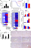
Figure 1.. Il20−/− mice are resistant to ConA-mediated acute hepatitis by elevating hepatoprotective cytokine IL-6 in the liver.
(A) C57BL/6N mice (n=6) were intravenously injected with ConA (12 mg/kg). Serum IL-20 levels were measured. (B-H) WT (n=8) and Il20−/− mice (n=10) were intravenously injected with ConA. (B) RT-qPCR of IL-20 mRNA levels. (C) Serum ALT and AST levels. (D) Representative images of H&E staining are shown, the percentage of necrotic area per field was quantified. (E, F) Serum cytokine levels and hepatic cytokine mRNA levels. (G, H) Western blot analyses of IL-6 and IFN-γ downstream signaling pathways, and pro- and anti-apoptotic protein levels in the liver post ConA injection. Values represent means ± SD. *P< 0.05, **P< 0.01, ***P< 0.001. ConA: Concanavalin A.
Figure 2.
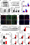
Figure 2.. Il20−/− mice have greater IL-6 protein expression in hepatocytes than WT mice in acute hepatitis.
(A-C) C57BL/6N mice (n=4–6) were intravenously injected with ConA (12 mg/kg). (A) RT-qPCR analyses of liver mRNA levels. (B) Western analyses and quantification of IL-20 receptors. (C) Representative images of immunohistochemistry staining of IL-20 receptors. (D, E) WT (n=4–8) and Il20−/− mice (n=4–8) were intravenously injected with ConA. (D) Representative images of immunofluorescence staining of IL-6. (E) Mean IL-6 fluorescence intensity was quantified. (F) Il6Hep−/− (n=8) and littermate Il6f/f control mice (n=7) were intravenously injected with ConA (12 mg/kg). Serum IL-6 levels were measured. Values represent means ± SD. *P< 0.05, **P< 0.01, ***P< 0.001. ConA: Concanavalin A.
Figure 3.
Figure 3.. Il20 deletion selectively enhances the expression of IκBζ-dependent genes including Il6 and Lcn2 in hepatocytes in acute hepatitis.
I (A-C) WT (n=4–8) and Il20−/− mice (n=4–8) were injected with ConA (12 mg/kg). (A) Western blot analyses of liver IκBζ expression. (B, C) RT-qPCR analyses of liver Nfkbiz mRNA and IκBζ-target genes. (D-F) C57BL/6N mice were injected with Ad-Gfp (n=3 in control group, n=5 in ConA-treated group) and Ad-shNfkbiz (n=3 in control group, n=6 in ConA-treated group) for 7 days, and followed by ConA injection. (D) RT-qPCR analyses of Nfkbiz mRNA. (E) Serum ALT and IL-6 levels were determined. (F) RT-qPCR analyses of liver IκBζ-target genes. Values represent means ± SD. *P< 0.05, **P< 0.01, ***P< 0.001. ConA: Concanavalin A
Figure 4.
Figure 4.. IL-20 deletion ameliorates bacterial infection by elevating hepatic expression of IκBζ-target antibacterial genes (eg. Il6 and Lcn2).
(A) C57BL/6N mice (n=6) were infected with K.P. (3000 CFU). Serum IL-20 levels were measured. (B, C) WT (n=19) and Il20−/− mice (n=18) were infected with K.P.. (B) Survival rates were analyzed. *P< 0.05. (C) Blood bacterial load was measured. (D, E) WT (n=4–7) and Il20−/− mice (n=4–8) were infected with K.P.. (D) RT-qPCR analyses of hepatic expression of IκBζ-target genes and other antibacterial genes. P values (Il20−/− versus WT mice at 3-h time point) are indicated. (E) Serum IL-6, LCN2 and IL-22 were measured. (F, G) RT-qPCR analyses of hepatic expression of Il20r mRNAs and representative images of liver IL-20 receptor staining in K.P.-infected mice. Values represent means ± SD. *P< 0.05. K.P.: Klebsiella pneumoniae
Figure 5.
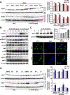
Figure 5.. IL-20 downregulates the expression of IκBζ protein and its target genes in hepatocytes by promoting IκBζ degradation.
(A, B) Schematic treatment timeline of mouse AML12 hepatocytes. (A) Western blot analysis and quantification of IκBζ expression. (B) RT-qPCR analyses of Nfkbiz and IκBζ-target gene mRNAs at the 3h time point. (C) Serum-starved AML12 cells were treated with IL-20, IL-1β, and/or various inhibitors as indicated. Western blot analysis and quantification of IκBζ expression. (D) Serum-starved AML12 cells were pretreated with IL-20 for 1h, and then stimulated with IL-1β in the presence or absence of MG132 for 2 h. Ubiquitination assay for IκBζ was detected with anti-ubiquitin. Values represent means ± SEM from three to four independent experiments. *P< 0.05, **P< 0.01, ***P< 0.001. K.P.: Klebsiella pneumoniae. IL-20 (50 ng/ml); IL-1β (20 ng/ml); CHX (100 μg/ml); MG132 (20 μM)
Figure 6
Figure 6. IL-20 promotes IκBζ degradation in hepatocytes via the induction of NQO1.
(A) Serum-starved AML12 cells or primary hepatocytes were treated with IL-20 (50 ng/ml) for the indicated time points. Western blot analysis and quantification of NQO1 expression. (B) AML12 cells were transfected with control siRNA or Nqo1 siRNA for 24 h, and then treated with IL-1β (20 ng/ml) for the indicated time points. Western blot analysis and quantification of IκBζ expression. (C, D) WT and Il20−/− mice were injected with ConA (12 mg/kg) or K.P. (3000 CFU) for the indicated time points. Liver tissues were subjected to the measurement of Nqo1 mRNA levels (panel C). Representative images of NQO1 (green), hepatocyte marker HNF-4α (red), and nuclei (blue) are shown in panel D. (E-H) C57BL/6N mice were intravenously injected with Ad-Gfp (n=3 in control group, n=6 in ConA-treated group) and Ad-shNqo1 (n=3 in control group, n=6 in ConA-treated group) for 7 days, and then followed by ConA injection. (E) Representative images of NQO1 (red) and nuclei (blue). (F) Western blot analysis and quantification of IκBζ expression. (G) Serum ALT and IL-6 levels. (H) RT-qPCR analyses of hepatic expression of IκBζ-dependent genes. Values represent means ± SEM from three to four independent in vitro experiments or means ± SD from in vivo experiments. *P< 0.05, **P< 0.01, ***P< 0.001.
Figure 7.
Figure 7.. IL-20 induces NQO1 expression in hepatocytes by activating ERK/NRF2 and p38/NRF2 signaling via the binding of IL-22R1/IL-20R2.
(A) Silencing of different IL-20 receptors in AML12 cells or primary hepatocytes was followed by serum-starvation for 1h, and treatment with IL-20 (50 ng/ml) for 4h. Western blot analysis and quantification of NQO1 expression. (B) Serum-starved primary hepatocytes were treated with IL-20 (100 ng/ml) or IL-22 (50 ng/ml) for the indicated time points. Western blot analyses of various proteins. (C) Serum-starved AML12 cells were treated with IL-20 (50 ng/ml) for the indicated time points. Western blot analysis and quantification of NRF2 expression (top panel). NRF2 nuclear translocation was analyzed by NRF2 immunofluorescence staining (lower panel). Representative images of NRF2 (green) and nuclei (blue) are shown. (D) Serum-starved AML12 cells or primary hepatocytes were pretreated with ERK1/2 inhibitor (PD98059, 50 μM), JNK inhibitor (SP600125, 50 μM), or p38 MAPK inhibitor (LY2228820, 2 μM) for 1h, and then stimulated with IL-20 (50 ng/ml) for 4h. Western blot analysis and quantification of NQO1 expression. Values represent means ± SEM from three to four independent experiments. *P< 0.05, **P< 0.01, ***P< 0.001.
Figure 8.
Figure 8.. Hepatic expression and correlation of IL1B, IL20, and IκBζ-target genes in SAH patients.
Human liver samples from healthy controls (HC, n=16), severe alcoholic hepatitis (SAH, n=20), and alcohol/HCV cirrhosis (Cirrhosis, n=28) were used. (A) RT-qPCR analyses. (B) A positive correlation of IL1B with IκBζ-target genes in human liver samples. (C) RT-qPCR analyses of IL-20 and NQO1 mRNA levels and a positive correlation of IL-20 with NQO1 in human liver samples. Values in panels A, C represent means ± SD. *P< 0.05, **P< 0.01, ***P< 0.001. (D) The schematic model depicting IL-20 exacerbates acute hepatitis and bacterial infection by downregulating IκBζ-target genes in hepatocytes. Under inflammatory conditions, inflammatory cells release various cytokines including IL-1β and IL-20. IL-1β induces IκBζ expression in hepatocytes and subsequently elevates its target genes including Il6 and Lcn2, which exert hepatoprotective function and limit bacterial infection. Meanwhile, IL-20 induces hepatic NQO1 expression by activating ERK/NRF2 and p38/NRF2 signaling pathway via the binding of IL-22R1/IL-20R2. The elevated NQO1 accelerates liver injury and bacterial infection by promoting IκBζ degradation and subsequent downregulation of IκBζ-target gene expression in hepatocytes.

Comment in

References

    1. Rutz S, Wang X, Ouyang W. The IL-20 subfamily of cytokines--from host defence to tissue homeostasis. Nat Rev Immunol 2014;14:783–795. - PubMed
    1. Sabat R, Ouyang W, Wolk K. Therapeutic opportunities of the IL-22-IL-22R1 system. Nat Rev Drug Discov 2014;13:21–38. - PubMed
    1. Arab JP, Sehrawat TS, Simonetto DA, Verma VK, Feng D, Tang T, et al. An Open-Label, Dose-Escalation Study to Assess the Safety and Efficacy of IL-22 Agonist F-652 in Patients With Alcohol-associated Hepatitis. Hepatology 2020;72:441–453. - PMC - PubMed
    1. Blumberg H, Conklin D, Xu WF, Grossmann A, Brender T, Carollo S, et al. Interleukin 20: discovery, receptor identification, and role in epidermal function. Cell 2001;104:9–19. - PubMed
    1. Chiu YS, Wei CC, Lin YJ, Hsu YH, Chang MS. IL-20 and IL-20R1 antibodies protect against liver fibrosis. Hepatology 2014;60:1003–1014. - PubMed

Publication types

MeSH terms