Review
. 2021 Jul 15;60(14):2315-2319.
doi: 10.2169/internalmedicine.6262-20. Epub 2021 Feb 22.

Cerebral Embolism Associated with Calcified Amorphous Tumor: A Review of Cerebral Infarction Cases

Affiliations
Review

Cerebral Embolism Associated with Calcified Amorphous Tumor: A Review of Cerebral Infarction Cases

Yamato Nishiguchi et al. Intern Med. .

Abstract

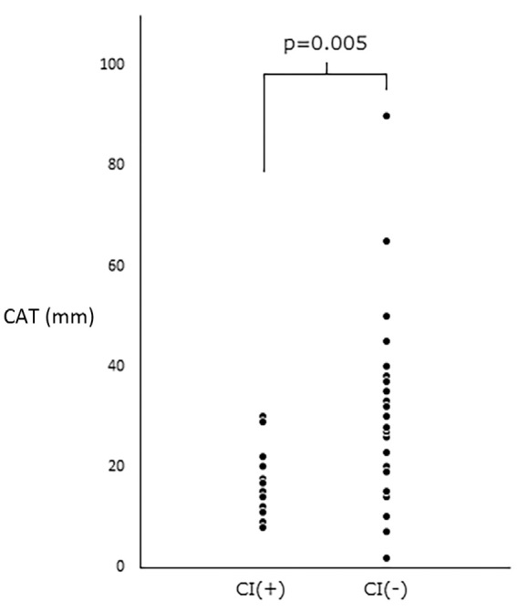
Calcified amorphous tumor (CAT) is a non-neoplastic tumor composed of calcified nodules consisting of amorphous fibrous material, and it may eventually cause cerebral infarction (CI). We experienced a 67-year-old woman with CAT who had recurrent CI. After excision of the CAT, the CI did not show recurrence. A review of previous papers on CI due to CAT in Pubmed revealed that 7 of 13 studies originated in Japan and that CI can occur even with small CAT. Surgical treatment is recommended to prevent CI recurrence, especially when CAT is accompanied by mitral annular calcification or has marked mobility.

Keywords: calcified amorphous tumor; cerebral infarction; embolization; mitral annular calcification.


Conflict of interest statement

The authors state that they have no Conflict of Interest (COI).

Figures

Figure 1.
Figure 1.
Radiological and ultrasonography findings. (A) Diffusion weighted image showed multiple cerebral infarctions in bilateral occipital lobes and cerebellum. (B) Magnetic resonance angiography showed a decreased blood flow in the left posterior cerebral artery (arrow), and the vertebral artery was right-dominant. (C) Contrast-enhanced computed tomography showed a high-density nodule in the mitral valve. (D) Three-dimensional computed tomography angiography showed left vertebral artery stenosis at the C5/6 level (arrow). (E) A transthoracic echocardiography examination revealed mitral annular calcification (arrow). (F) Transesophageal echocardiography showed swinging calcified amorphous tumor (arrow).
Figure 2.
Figure 2.
Pathological findings. (A) The gross anatomic pathology of the excised tumor is yellow-white in color and partially calcified. (B) A histological examination revealed multiple nodular calcifications surrounded by fibrous connective tissue and fibrin. There was no identifiable myxoma or inflammatory cell infiltration (Hematoxylin and Eosin staining; bar=500 μm).
Figure 3.
Figure 3.
The maximum length of the CAT (mm). The CAT that caused CI was significantly shorter than the CAT without CI. CAT: calcified amorphous tumor, CI: cerebral infarction

References

    1. Reynolds C, Tazelaar HD, Edwards WD. Calcified amorphous tumor of the heart (cardiac CAT). Hum Pathol 28: 601-606, 1997. - PubMed
    1. Elbardissi AW, Dearani JA, Daly RC, et al. . Survival after resection of primary cardiac tumors: a 48-year experience. Circulation 118(Suppl): S7-S15, 2008. - PubMed
    1. Watanabe Y, Naganuma T, Nakao T, Nakamura S. A calcified amorphous tumor originating in the sinus of valsalva. Echocardiography 33: 796-798, 2016. - PubMed
    1. de Hemptinne Q, de Cannière D, Vandenbossche JL, Unger P. Cardiac calcified amorphous tumor: a systematic review of the literature. Int J Cardiol Heart Vasc 7: 1-5, 2015. - PMC - PubMed
    1. Tsuchihashi K, Nozawa A, Marusaki S, et al. . Mobile intracardiac calcinosis: a new risk of thromboembolism in patients with haemodialysed end stage renal disease. Heart 82: 638-640, 1999. - PMC - PubMed