Review
. 2021 Feb 22;6(1):79.
doi: 10.1038/s41392-020-00455-6.

A double-edged sword of immuno-microenvironment in cardiac homeostasis and injury repair

Affiliations
Review

A double-edged sword of immuno-microenvironment in cardiac homeostasis and injury repair

Kang Sun et al. Signal Transduct Target Ther. .

Abstract

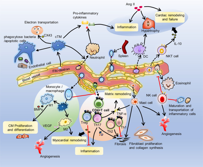
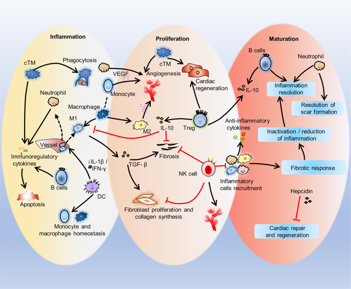
The response of immune cells in cardiac injury is divided into three continuous phases: inflammation, proliferation and maturation. The kinetics of the inflammatory and proliferation phases directly influence the tissue repair. In cardiac homeostasis, cardiac tissue resident macrophages (cTMs) phagocytose bacteria and apoptotic cells. Meanwhile, NK cells prevent the maturation and transport of inflammatory cells. After cardiac injury, cTMs phagocytose the dead cardiomyocytes (CMs), regulate the proliferation and angiogenesis of cardiac progenitor cells. NK cells prevent the cardiac fibrosis, and promote vascularization and angiogenesis. Type 1 macrophages trigger the cardioprotective responses and promote tissue fibrosis in the early stage. Reversely, type 2 macrophages promote cardiac remodeling and angiogenesis in the late stage. Circulating macrophages and neutrophils firstly lead to chronic inflammation by secreting proinflammatory cytokines, and then release anti-inflammatory cytokines and growth factors, which regulate cardiac remodeling. In this process, dendritic cells (DCs) mediate the regulation of monocyte and macrophage recruitment. Recruited eosinophils and Mast cells (MCs) release some mediators which contribute to coronary vasoconstriction, leukocyte recruitment, formation of new blood vessels, scar formation. In adaptive immunity, effector T cells, especially Th17 cells, lead to the pathogenesis of cardiac fibrosis, including the distal fibrosis and scar formation. CMs protectors, Treg cells, inhibit reduce the inflammatory response, then directly trigger the regeneration of local progenitor cell via IL-10. B cells reduce myocardial injury by preserving cardiac function during the resolution of inflammation.


Conflict of interest statement

The authors declare no competing interests.

Figures

Fig. 1
Fig. 1
Types of cardiac tissue injury. Ischemia-reperfusion injury (IRI), permanent ligation injury (PLI), cryoinjury, resection, and gene ablation are the four main ways of studying cardiac injury and repair. IRI is used to simulate the pathological state of myocardial infarction (MI). The ligation is untied and blood reperfusion is performed after ligation of the mouse artery for 30 min Cryoinjury is caused by cauterization of ventricular tissue with a cryoprobe. Resection is used to surgically remove part of the cardiac tissues. The gene ablation method specifically expresses bacterial nitroreductase (NTR) or diphtheria toxin receptor (DTR) in cardiomyocytes (CMs) to result in CM death
Fig. 2
Fig. 2
Molecular therapy of injured cardiac tissues. Some molecules promote repair (blue line), while others inhibit repair (red line) after cardiac injury. Growth factors such as VEGFA, FGF2, and NRG1, the NRG1 receptors ERBB2 and ERBB4, exosomes, and miR-199a or miR-590 restore cardiac function. miR-199a or miR-590, the miR-17-92 cluster, miR-214, miR-302-367, and miR-222 induce CM proliferation. The miR-17-92 cluster, miR-214, miR-302-367, miR-222 and miR-199a or miR-590 reduce fibrosis. FGF2 and exosomes reduce infarct size. Exosomes, Nrf2 and Pitx2 scavenge ROS. Pitx2 also regulates electron transport. Exosomes can act as mediators of cell-to-cell communication. NRG1 and its receptors ERBB2 and ERBB4 and VEGFA improve vascular regeneration. VEGFA also improves local coronary blood flow
Fig. 3
Fig. 3
Cell therapy of injured cardiac tissues. iPSCs can differentiate into functional cardiomyocytes in vitro. Heart-derived cells can differentiate into many types of heart cells. GMT or GHMT mixtures reprogram mouse fibroblasts to differentiate into iCMs, and Smarcd3 reprograms mouse fibroblasts to differentiate into cardiac progenitor cells in vitro. MSCs differentiate into CMs in the presence of 5-aza or in coculture with cardiac progenitor cells. However, reprogramming human fibroblasts requires GMT or GHMT mixtures and troponin, MESP1, ESRRγ and ZFPM2, was well as miR-1 and miR-133. Human fibroblasts can be transformed into cardiac progenitor cells with c-ETS2 and MESP1
Fig. 4
Fig. 4
Immune cells in the heart. cTMs internalize blood-borne FITC-dextran; cTMs also have typical macrophage characteristics and phagocytose bacteria and apoptotic cells. cTMs regulate electrical conduction through CX43, and cTMs can also produce proinflammatory cytokines to induce inflammation in the aging heart and promote neutrophil infiltration. Resident NK cells reduce cardiac eosinophil infiltration. NK cells prevent the maturation and transport of inflammatory cells. Monocytes are recruited into cardiac tissue and differentiate into M1 and M2 macrophages. M1 macrophages affect the proliferation and differentiation of CMs by BMPs, and M2 macrophages are related to angiogenesis by producing VEGF. Recruited neutrophils secrete proinflammatory cytokines, which promote fibroblast differentiation and lead to sustained inflammation. DCs upregulate cardiomyocyte hypertrophy a. NKT cells secrete cytokines such as IL-10 to protect or regulate hypertrophy, cardiac remodeling and the inflammatory response induced by Ang II. Infiltrating CD4 + cells include helper T cells (Th1 and Th2), Th17 cells and Treg cells. Th1 cells reduce the fibrotic response, while Th2 and Th17 cells promote fibrosis. Th17 cells also promote inflammation and extracellular matrix remodeling, and Treg cells reduce inflammation. Mast cells promote fibrosis, angiogenesis and cardiac fibroblast proliferation and collagen synthesis through TNF-α
Fig. 5
Fig. 5
Three phases of immune response to cardiac injury repair. This diagram shows the inflammation, proliferation, and maturation phases after cardiac injury. In the Inflammatory phase, cTMs phagocytose dying cardiomyocytes. B cells secrete immunoregulatory factors to reduce cardiac contractility and promote cardiomyocyte apoptosis. DCs mediate the recruitment of inflammatory cells such as monocytes and M1 macrophages and homeostasis. In the proliferation phase, cTMs promote the proliferation of myocardial cells and angiogenesis. Neutrophils, monocytes and M2 macrophages also promote angiogenesis though VEGF, and M1 macrophages promote tissue fibrosis and myocardial remodeling by inducing extracellular matrix release from cardiac fibroblasts. NK cells protect against cardiac fibrosis by directly restricting collagen formation of cardiac fibroblasts and preventing the accumulation of specific inflammatory populations, and NK cells also promote blood vessel remodeling. Treg cells inhibit inflammation and fibrosis and promote precursor cell proliferation and macrophage polarization. In the maturation phase, the recruitment of inflammatory cells such as macrophages, neutrophils and eosinophils is inhibited, anti-inflammatory cytokines are secreted, infiltrating immune cells regulate inflammation inactivation/reduction by mediating the fibrotic response, and inflammation and scar formation are resolved. During this phase, hepcidin inhibits macrophage-induced cardiac repair and regeneration

Similar articles

Cited by

References

    1. Ma X, et al. An exceptionally preserved arthropod cardiovascular system from the early Cambrian. Nat. Commun. 2014;5:3560. doi: 10.1038/ncomms4560. - DOI - PubMed
    1. Gosavi S, Tyroch AH, Mukherjee D. Cardiac trauma. Angiology. 2016;67:896–901. doi: 10.1177/0003319715627954. - DOI - PubMed
    1. Lozano R, et al. Global and regional mortality from 235 causes of death for 20 age groups in 1990 and 2010: a systematic analysis for the Global Burden of Disease Study 2010. Lancet. 2012;380:2095–2128. doi: 10.1016/S0140-6736(12)61728-0. - DOI - PMC - PubMed
    1. Godwin JW, Pinto AR, Rosenthal NA. Macrophages are required for adult salamander limb regeneration. Proc. Natl. Acad. Sci. USA. 2013;110:9415–9420. doi: 10.1073/pnas.1300290110. - DOI - PMC - PubMed
    1. Aurora AB, et al. Macrophages are required for neonatal heart regeneration. J. Clin. Investig. 2014;124:1382–1392. doi: 10.1172/JCI72181. - DOI - PMC - PubMed

Publication types

MeSH terms