. 2021;31(2):145-147.
doi: 10.1007/s00194-021-00462-z. Epub 2021 Feb 15.

COVID-19 lungs in post-mortem computed tomography

Affiliations

COVID-19 lungs in post-mortem computed tomography

I Kniep et al. Rechtsmedizin (Berl). 2021.

Abstract

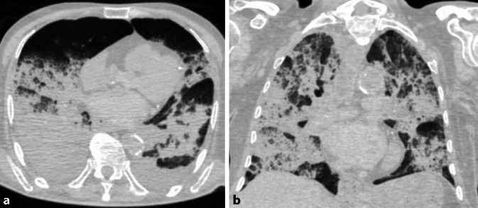
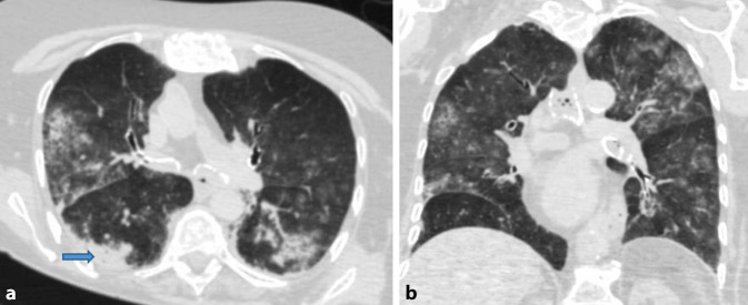
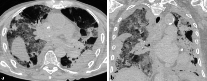
Post-mortem computed tomography (PMCT) is a rapid and noninvasive diagnostic tool for important contributions to the identification of pulmonary findings in the deceased with pneumonia, including cases of coronavirus disease 2019 (COVID-19). Although computed tomography (CT) shows a high sensitivity for pneumonia in living persons, it is relatively unspecific for COVID-19 pneumonia clinically. Typical CT findings for viral pneumonia therefore require confirmation by PCR tests (polymerase chain reaction tests), even if lung infections with severe acute respiratory syndrome coronavirus type 2 (SARS-CoV-2) show characteristic patterns, most frequently ground glass opacities (GGO) and a combination of GGO and air space consolidations. In the consecutive autopsy series of SARS-CoV‑2 deaths from Hamburg, Germany, the most frequent cause of death was and still is COVID-19 pneumonia. Typical findings were frequently found in the PMCT in SARS-CoV-2-associated deaths, which were taken into account when classifying the death as COVID-19.

Die Post-mortem-Computertomographie (PMCT) ist ein schnelles und nicht-invasives Diagnoseinstrument, das einen wichtigen Beitrag zur Identifizierung von Lungenbefunden bei Verstorbenen mit Pneumonie leistet – auch bei Fällen der Coronaviruskrankheit (COVID -19). Obwohl die Computertomografie (CT) eine hohe Sensitivität für Lungenentzündungen bei lebenden Personen aufweist, ist sie für eine COVID-19-Pneumonie klinisch recht unspezifisch. Typische CT-Befunde für virale Pneumonien bedürfen daher der Bestätigung durch PCR-Tests (Polymerase-Ketten-Reaktion-Tests), auch wenn die Lungeninfektion mit SARS-CoV‑2 charakteristische Muster zeigt – am häufigsten Milchglastrübungen (GGO) und eine Kombination aus GGO und Konsolidierungen.In der konsekutiven Autopsieserie von SARS-CoV-2-Todesfällen aus Hamburg war und ist die häufigste Todesursache die COVID-19-Pneumonie. In der PMCT fanden sich bei SARS-CoV-2-assoziierten Todesfällen häufig typische Befunde, die bei der Klassifizierung des Todes als COVID-19 berücksichtigt wurden.

Keywords: Consolidation; Ground glass opacities; Hypostasis; Pulmonary manifestation; SARS-CoV-2.


Conflict of interest statement

Conflict of interestI. Kniep, A. Heinemann, C. Edler, J.P. Sperhake, K. Püschel, B. Ondruschka and A.S. Schröder declare that they have no competing interests.

Figures

Fig. 1
Fig. 1
Post-mortem computed tomography case 1. a Axial CT image (lung section). b Coronary CT image (lung section)
Fig. 2
Fig. 2
Post-mortem computed tomography case 2. a Axial CT image (lung section; blue arrow directing to the area of consolidation). b Coronary CT image (lung section)
Fig. 3
Fig. 3
Post-mortem computed tomography case 3. a Axial CT image (section of the lung). b Coronary CT image (lung section)

Similar articles

Cited by

  • Technologies for Studying and Teaching Human Anatomy: Implications in Academic Education.
    Graziani GC, Bocchi M, Gouvêa-E-Silva LF, Fornaziero CC, Fernandes EV. Graziani GC, et al. Med Sci Educ. 2024 May 28;34(5):1203-1214. doi: 10.1007/s40670-024-02079-9. eCollection 2024 Oct. Med Sci Educ. 2024. PMID: 39450022 Review.
  • Postmortem CT in decedents with SARS-CoV-2 infection. A single institution experience.
    Thomas M, Abtin F, Roth A, Yim C, Pahwa A, Paige J, Ukpo O. Thomas M, et al. Forensic Sci Res. 2022 Jan 24;7(2):255-260. doi: 10.1080/20961790.2021.1977479. eCollection 2022. Forensic Sci Res. 2022. PMID: 35784405 Free PMC article.
  • Postmortem Chest Computed Tomography in Fatal COVID-19: A Valuable Diagnostic Tool for Minimally Invasive Autopsy.
    Silva PSDD, Sawamura MVY, Monteiro RAA, Duarte-Neto AN, Martin MDGM, Dolhnikoff M, Mauad T, Saldiva PHN, Leite CC, Silva LFFD, Cardoso EF. Silva PSDD, et al. Clinics (Sao Paulo). 2021 Dec 8;76:e3551. doi: 10.6061/clinics/2021/e3551. eCollection 2021. Clinics (Sao Paulo). 2021. PMID: 34909914 Free PMC article. No abstract available.
  • Organ manifestations of COVID-19: what have we learned so far (not only) from autopsies?
    Jonigk D, Werlein C, Acker T, Aepfelbacher M, Amann KU, Baretton G, Barth P, Bohle RM, Büttner A, Büttner R, Dettmeyer R, Eichhorn P, Elezkurtaj S, Esposito I, Evert K, Evert M, Fend F, Gaßler N, Gattenlöhner S, Glatzel M, Göbel H, Gradhand E, Hansen T, Hartmann A, Heinemann A, Heppner FL, Hilsenbeck J, Horst D, Kamp JC, Mall G, Märkl B, Ondruschka B, Pablik J, Pfefferle S, Quaas A, Radbruch H, Röcken C, Rosenwald A, Roth W, Rudelius M, Schirmacher P, Slotta-Huspenina J, Smith K, Sommer L, Stock K, Ströbel P, Strobl S, Titze U, Weirich G, Weis J, Werner M, Wickenhauser C, Wiech T, Wild P, Welte T, von Stillfried S, Boor P. Jonigk D, et al. Virchows Arch. 2022 Aug;481(2):139-159. doi: 10.1007/s00428-022-03319-2. Epub 2022 Apr 1. Virchows Arch. 2022. PMID: 35364700 Free PMC article. Review.
  • Use of post-mortem chest computed tomography in Covid-19 pneumonia.
    De-Giorgio F, Cittadini F, Cina A, Cavarretta E, Biondi-Zoccai G, Vetrugno G, Natale L, Colosimo C, Pascali VL. De-Giorgio F, et al. Forensic Sci Int. 2021 Aug;325:110851. doi: 10.1016/j.forsciint.2021.110851. Epub 2021 May 27. Forensic Sci Int. 2021. PMID: 34090259 Free PMC article.

References

    1. Carotti M, Salaffi F, Sarzi-Puttini P, Agostini A, Borgheresi A, Minorati D, Galli M, Marotto D, Giovagnoni A. Chest CT features of coronavirus disease 2019 (COVID-19) pneumonia: key points for radiologists. Radiol Med. 2020;125(7):636–646. doi: 10.1007/s11547-020-01237-4. - DOI - PMC - PubMed
    1. Christie A, Flach P, Ross S, Spendlove D, Bolliger S, Vock P, Thali MJ. Clinical radiology and postmortem imaging (virtopsy) are not the same: specific and unspecific postmortem signs. Leg Med. 2010;12(5):215–222. doi: 10.1016/j.legalmed.2010.05.005. - DOI - PubMed
    1. Chung M, Bernheim A, Mei X, Zhang N, Huang M, Zeng X, Cui J, Xu W, Yang Y, Fayad ZA, Jacobi A, Li K, Li S, Shan H. CT imaging features of 2019 novel coronavirus (2019-nCoV) Radiology. 2020;295(1):202–207. doi: 10.1148/radiol.2020200230. - DOI - PMC - PubMed
    1. Ducloyer M, Gaborit B, Toquet C, Castain L, Bal A, Arrigoni PP, Lecomte R, Clement R, Sagan C. Complete post-mortem data in a fatal case of COVID-19: clinical, radiological and pathological correlations. Int J Legal Med. 2020 doi: 10.1007/s00414-020-02390-1. - DOI - PMC - PubMed
    1. Edler C, Schröder AS, Aepfelbacher M, Fitzek A, Heinemann A, Heinrich F, Klein A, et al. Dying with SARS-CoV-2 infection—an autopsy study of the first consecutive 80 cases in Hamburg, Germany. Int J Legal Med. 2020;134(4):1275–1284. doi: 10.1007/s00414-020-02317-w. - DOI - PMC - PubMed

LinkOut - more resources