Review
. 2021 Feb 5:15:630016.
doi: 10.3389/fnins.2021.630016. eCollection 2021.

Selenium at the N eural B arriers: A R eview

Affiliations
Review

Selenium at the N eural B arriers: A R eview

Nikolay Solovyev et al. Front Neurosci. .

Abstract

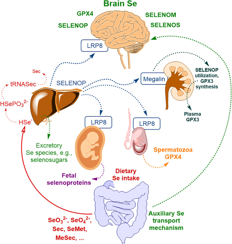
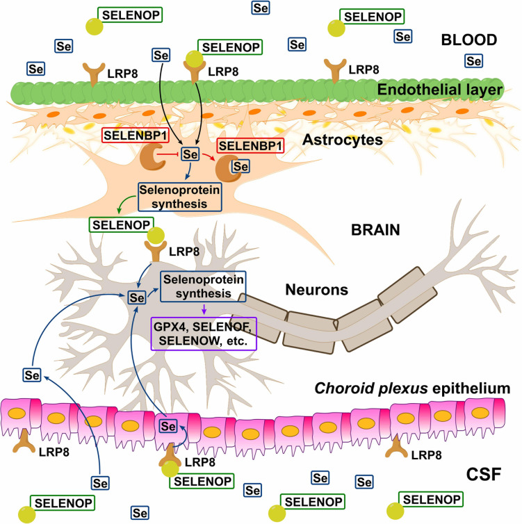
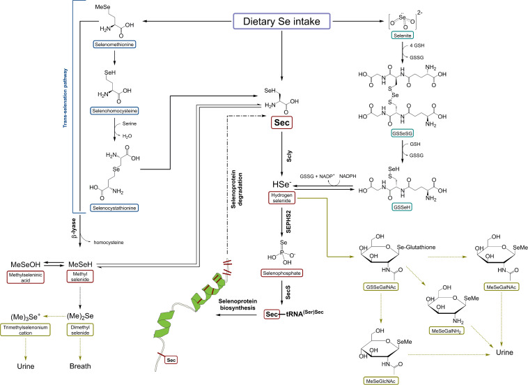
Selenium (Se) is known to contribute to several vital physiological functions in mammals: antioxidant defense, fertility, thyroid hormone metabolism, and immune response. Growing evidence indicates the crucial role of Se and Se-containing selenoproteins in the brain and brain function. As for the other essential trace elements, dietary Se needs to reach effective concentrations in the central nervous system (CNS) to exert its functions. To do so, Se-species have to cross the blood-brain barrier (BBB) and/or blood-cerebrospinal fluid barrier (BCB) of the choroid plexus. The main interface between the general circulation of the body and the CNS is the BBB. Endothelial cells of brain capillaries forming the so-called tight junctions are the primary anatomic units of the BBB, mainly responsible for barrier function. The current review focuses on Se transport to the brain, primarily including selenoprotein P/low-density lipoprotein receptor-related protein 8 (LRP8, also known as apolipoprotein E receptor-2) dependent pathway, and supplementary transport routes of Se into the brain via low molecular weight Se-species. Additionally, the potential role of Se and selenoproteins in the BBB, BCB, and neurovascular unit (NVU) is discussed. Finally, the perspectives regarding investigating the role of Se and selenoproteins in the gut-brain axis are outlined.

Keywords: LRP8; blood–brain barrier; blood–cerebrospinal fluid barrier; brain-gut axis; low molecular weight selenium species; selenium; selenium transport; selenoprotein P.


Conflict of interest statement

The authors declare that the research was conducted in the absence of any commercial or financial relationships that could be construed as a potential conflict of interest.

Figures

FIGURE 1
FIGURE 1
The scheme of body Se homeostasis. Abbreviations: LRP8 – low-density lipoprotein receptor-related protein 8 (LRP8, also known as apolipoprotein E receptor-2, ApoER2), GPX3 – glutathione peroxidase type III, GPX4 – glutathione peroxidase type IV, Sec – selenocysteine, MeSec – methyl selenocysteine, SELENOM – selenoprotein M, SELENOP – selenoprotein P, SELENOS – selenoprotein S; * – auxiliary brain Se transport mechanism, independent of SELENOP, possibly related to selenosugars (Burk and Hill, 2015) and other low molecular weight Se-species (Solovyev et al., 2013) and possibly other minor contributors (please, see text for more detail). Based on Solovyev et al. (2018) with modification.
FIGURE 2
FIGURE 2
Hypothetical model of Se transport across blood–brain barrier (BBB) and blood–cerebrospinal fluid barrier (BCB). Circulating SELENOP present in blood and CSF is taken up by LRP8-positive cells in the epithelial (BBB) and ependymal (BCB) layers, resynthesized in neighboring astrocytes, and released to supply LRP8-positive neurons with Se. In the astrocytes, SELENBP1 sequesters Se from selenoprotein synthesis and thus negatively regulating SELENOP production. There is also evidence indicating the existence of the SELENOP-independent Se uptake pathway (Figure 1). Reproduced from Sasuclark et al. (2019) with modification.
FIGURE 3
FIGURE 3
A schematic representation of selenocysteine β-lyase role in Se metabolism. Scly – selenocysteine β-lyase; Sec – selenocysteine; SeMet – selenomethionine; HSe – hydrogen selenide; GSSeSG – selenodiglutathione; GSSeH – selenoglutathione; SEPHS2 – selenophosphate synthetase 2. Based on Seale (2019) with modification.

References

    1. Abbott N. J., Patabendige A. A. K., Dolman D. E. M., Yusof S. R., Begley D. J. (2010). Structure and function of the blood–brain barrier. Neurobiol. Dis. 37 13–25. 10.1016/j.nbd.2009.07.030 - DOI - PubMed
    1. Adriani G., Ma D., Pavesi A., Kamm R. D., Goh E. L. (2017). A 3D neurovascular microfluidic model consisting of neurons, astrocytes and cerebral endothelial cells as a blood-brain barrier. Lab. Chip. 17 448–459. 10.1039/c6lc00638h - DOI - PubMed
    1. Aguilar M. V., Jiménez-Jiménez F. J., Molina J. A., Meseguer I., Mateos-Vega C. J., González-Muñoz M. J., et al. (1998). Cerebrospinal fluid selenium and chromium levels in patients with Parkinson’s disease. J. Neural. Transm. 105 1245–1251. 10.1007/s007020050127 - DOI - PubMed
    1. Ahn S. I., Sei Y. J., Park H.-J., Kim J., Ryu Y., Choi J. J., et al. (2020). Microengineered human blood–brain barrier platform for understanding nanoparticle transport mechanisms. Nat. Commun. 11:175. 10.1038/s41467-019-13896-7 - DOI - PMC - PubMed
    1. Arner E. S. (2010). Selenoproteins-What unique properties can arise with selenocysteine in place of cysteine? Exp. Cell Res. 316 1296–1303. 10.1016/j.yexcr.2010.02.032 - DOI - PubMed