Review
. 2021 Feb 3:14:610857.
doi: 10.3389/fnmol.2021.610857. eCollection 2021.

Rapid and Reversible Development of Axonal Varicosities: A New Form of Neural Plasticity

Affiliations
Review

Rapid and Reversible Development of Axonal Varicosities: A New Form of Neural Plasticity

Chen Gu. Front Mol Neurosci. .

Abstract

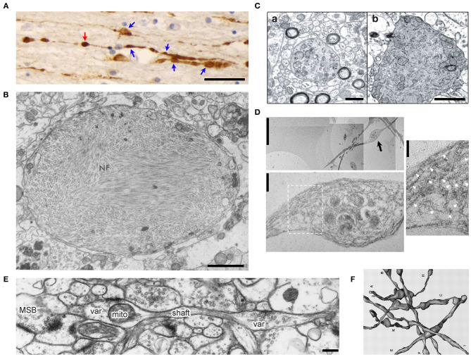
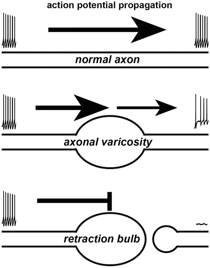
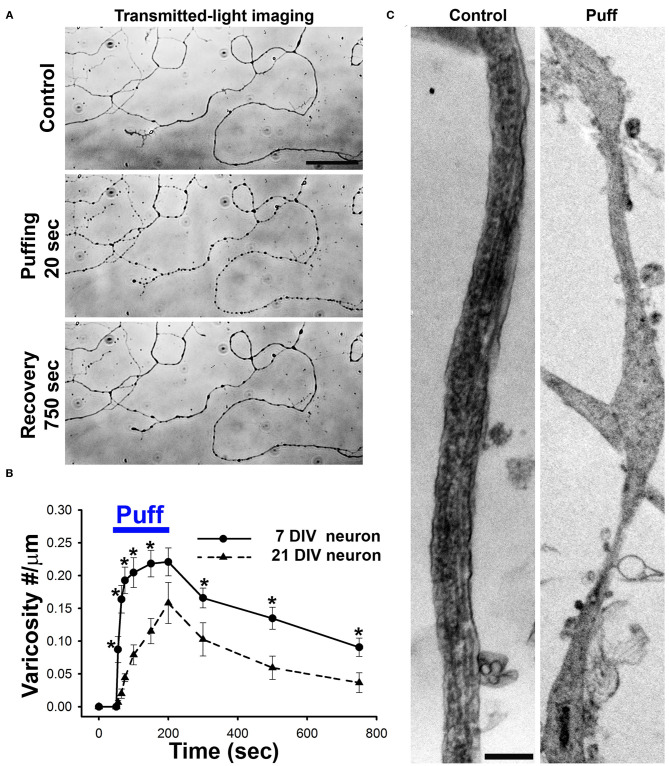
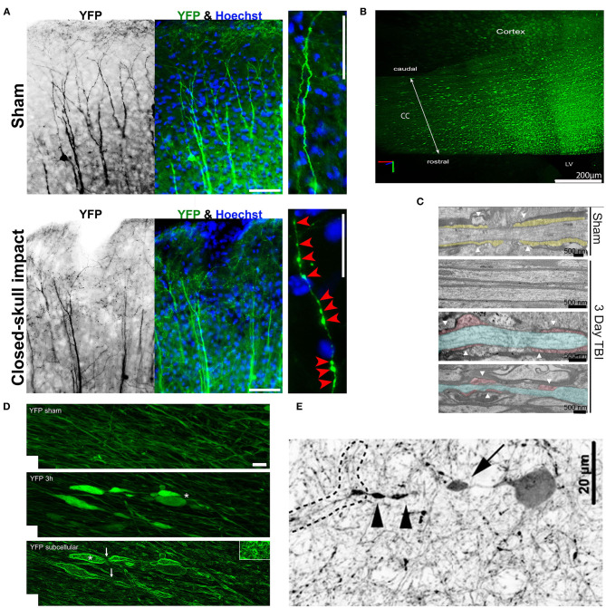
Axonal varicosities are enlarged, heterogeneous structures along axonal shafts, profoundly affecting axonal conduction and synaptic transmission. They represent a key pathological feature believed to develop via slow accumulation of axonal damage that occurs during irreversible degeneration, for example in mild traumatic brain injury (mTBI), Alzheimer's and Parkinson's diseases, and multiple sclerosis. Here this review first discusses recent in vitro results showing that axonal varicosities can be rapidly and reversibly induced by mechanical stress in cultured primary neurons from the central nervous system (CNS). This notion is further supported by in vivo studies revealing the induction of axonal varicosities across various brain regions in different mTBI mouse models, as a prominent feature of axonal pathology. Limited progress in understanding intrinsic and extrinsic regulatory mechanisms of axonal varicosity induction and development is further highlighted. Rapid and reversible formation of axonal varicosities likely plays a key role in CNS neuron mechanosensation and is a new form of neural plasticity. Future investigation in this emerging research field may reveal how to reverse axonal injury, contributing to the development of new strategies for treating brain injuries and related neurodegenerative diseases.

Keywords: Alzheimer's disease; action potential; axonal varicosity; mechanosensitive ion channel; microtubule; mild traumatic brain injury; neural plasticity; synaptic transmission.


Conflict of interest statement

The author declares that the research was conducted in the absence of any commercial or financial relationships that could be construed as a potential conflict of interest.

Figures

Figure 1
Figure 1
Morphology of axonal varicosities in injured, diseased or normal brains. (A), Post-mortem APP immunoreactivity within the corpus callosum of a TBI patient displayed multiple varicosities (blue arrows) along individual axons (Tang-Schomer et al., 2012). The swelling without a visible thin linking axon is a potential terminal bulb (red arrow). Scale bar, 30 μm. (B), The transmission electron microscope (TEM) image of an axonal varicosity or swelling filled with neurofibrils (NF) from the post-mortem cerebral cortex of a patient with Alzheimer's presenile dementia (Terry et al., 1964). Scale bar, 1 μm. (C), Ultrastructure of axonal varicosity in a mouse model of AD (Stokin et al., 2005). Degenerative changes in axonal varicosity along the fibers of the nucleus basalis of Meynert in Tg-swAPPPrp (b) but not wild-type mice (a). Scale bar, 1 μm. (D), TEM images of cultured cortical neurons from spastin−/− mice show disorganization of the microtubule (MT) network within axonal varicosities (Fassier et al., 2013). The image in the lower panel is the high magnification of axonal varicosity indicated by a blackarrow in the upper image. The image on the right is the high magnification of boxed regions, showing the tangled and bent aspect of MT filaments within the axonal varicosity. This abnormal appearance of MTs was never observed in wild-type axons, in which MTs were always organized in parallel arrays. White arrows indicate MT. Scale bars: 5 μm (upper left), 0.5 μm (lower left) and 0.25 μm (right). (E), Ultrastructural features of CA3 varicosities (Var) and axons in stratum radiatum of area CA1 in the normal rat brain (Shepherd and Harris, 1998). Longitudinally sectioned axon with two boutons, neither of which had additional PSDs or mitochondria in adjacent images. mito, mitochondria; MSB, multiple-synapse bouton; PSD, postsynaptic density. Scale bar, 1 μm. (F), Eight reconstructed axons from series electron microscopy images including axonal varicosities (Shepherd and Harris, 1998).
Figure 2
Figure 2
Effects of an axonal varicosity or a terminal bulb on action potential propagation along an axon. Action potentials propagate along a normal axon with high fidelity (top). Altered waveform, reduced frequency and even failure of action potentials can be induced by axonal varicosity (middle). A terminal bulb indicates a broken axon, which no longer allows action potentials to travel through.
Figure 3
Figure 3
Axonal varicosities are rapidly and reversibly induced by mechanical stress in cultured CNS neurons. (A), Puffing induced rapid and reversible formation of axonal varicosities in cultured hippocampal neurons at 7 days in vitro (DIV), revealed by microscopy with transmitted lights (Gu et al., 2017). Mechanical pressure was delivered by puffing of Hank's buffer via a glass pipette onto cultured neurons with 190 mm H2O static pressure at the tip for 150 s. Axonal varicosities slowly and partially recovered after puffing. (B), Summary of puffing-induced varicosity densities along axons from younger (7 DIV) and older (21 DIV) neurons (Gu et al., 2017). (C), TEM images of axonal segments under control condition (left) and when developing multiple varicosities induced by puffing (right) (Gu et al., 2017). Scale bars, 50 μm in (A) and 1 μm in (C).
Figure 4
Figure 4
Axonal varicosities induced in vivo by mechanical impact in mouse models of mTBI. (A), The repetitive closed-skull impact model induced axonal varicosities in multi-focal fashion in the cortex of Thy1-YFP transgenic mice (Gu et al., 2017). Mice were perfused and fixed immediately after the 2nd impact. Sham and impacted mice, upper and lower panels. YFP fluorescent signals are inverted in the gray scale image on the left. The apical dendrites of layer V projection neurons in the cortex point in the upward direction. Higher magnification images on the right show individual axons. Red arrowheads, induced axonal varicosities. Scale bars: 100 μm for left/middle panels and 20 μm for right panels. (B), Concussive TBI (closed-skull) induced YFP+ axonal varicosities in the corpus callosum (CC) under the impact site illustrated using CLARITY (Marion et al., 2018). The mouse was sacrificed 3 days after impact. LV, lateral ventricle. (C), TEM images of coronal section through the CC to illustrate organized myelin loop attachments forming paranodes (yellow fill; white arrowheads) in the sham (top), and axons in impacted mice with normal myelin and adjacent damaged axons (blue) with cytoskeletal breakdown paranodes (red fill) and myelin loss (bottom) (Marion et al., 2018). (D), Axonal varicosities were induced in the corticospinal tract 3 h after weight-drop impact (Ziogas and Koliatsos, 2018). Confocal images of sham (top) and injured (middle with conventional confocal microscopy and bottom using edge-detection function. White asterisks, axonal terminal bulbs; White arrows, thin axons bridging swellings. Scale bar, 5 μm. (E), A representative tdTomato+ interneuron showing a perisomatic axonal varicosity (arrow) and more distal varicosities (arrowheads) induced in a central fluid percussion model (Vascak et al., 2018).

Similar articles

Cited by

References

    1. Abrahams S., Mc Fie S., Patricios J., Suter J., September A. V., Posthumus M. (2019). Toxic tau: the TAU gene polymorphisms associate with concussion history in rugby union players. J. Sci. Med. Sport. 22, 22–28. 10.1016/j.jsams.2018.06.012 - DOI - PubMed
    1. Amack J. D., Manning M. L. (2012). Knowing the boundaries: extending the differential adhesion hypothesis in embryonic cell sorting. Science 338, 212–215. 10.1126/science.1223953 - DOI - PubMed
    1. Antic S., Wuskell J. P., Loew L., Zecevic D. (2000). Functional profile of the giant metacerebral neuron of Helix aspersa: temporal and spatial dynamics of electrical activity in situ. J. Physiol. 527(Pt 1), 55–69. 10.1111/j.1469-7793.2000.00055.x - DOI - PMC - PubMed
    1. Arnadottir J., Chalfie M. (2010). Eukaryotic mechanosensitive channels. Annu. Rev. Biophys. 39, 111–137. 10.1146/annurev.biophys.37.032807.125836 - DOI - PubMed
    1. Barrientos S. A., Martinez N. W., Yoo S., Jara J. S., Zamorano S., Hetz C., et al. . (2011). Axonal degeneration is mediated by the mitochondrial permeability transition pore. J. Neurosci. 31, 966–978. 10.1523/JNEUROSCI.4065-10.2011 - DOI - PMC - PubMed

LinkOut - more resources