

Conjunctival Intraepithelial Neoplasia in a Patient Presenting with Pigmented Conjunctival Lesion
- PMID: 33613255
- PMCID: PMC7879333
- DOI: 10.1159/000510570
Conjunctival Intraepithelial Neoplasia in a Patient Presenting with Pigmented Conjunctival Lesion
Abstract
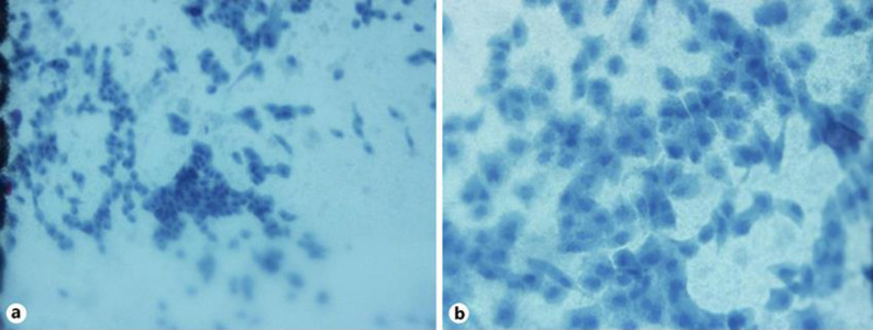
We report a case of conjunctival intraepithelial neoplasia (CIN) in a patient presenting with the pigmented conjunctival lesion. This study involved a 56-year-old woman that presented with right eye irritation for 1 month. She noticed brownish pigmentation arising from her right nasal conjunctiva and growing slowly over time. Biomicroscopic examination showed a gelatinous pigmented conjunctival mass with feeder vessels. Conjunctival impression cytology (CIC) was done and reported as CIN. Treatment was started with 0.02% mitomycin-C eye drops. The conjunctival lesion responded well to medication. This report shows that CIN can manifest as a pigmented tumor, resembling melanoma. CIC plays a role in the diagnosis of this condition. This tumor responded well with 0.02% mitomycin-C eye drops.
Keywords: Conjunctival intraepithelial neoplasia; Conjunctival melanoma; Pigmented conjunctival lesion.
Copyright © 2021 by S. Karger AG, Basel.
Conflict of interest statement
There are no conflicts of interest to report for all authors.
Figures


Similar articles
-
Conjunctival Intraepithelial Neoplasia Mimicking a Pigmentary Lesion in an HIV-Seropositive Indian Male.Cureus. 2024 Apr 24;16(4):e58953. doi: 10.7759/cureus.58953. eCollection 2024 Apr. Cureus. 2024. PMID: 38800191 Free PMC article.
-
[Treatment of 9 squamous epithelial carcinoma in situ lesions of the conjunctiva (CIN) with mitomycin C eyedrops in cytological and DNA image cytometric control].Klin Monbl Augenheilkd. 2001 Jun;218(6):429-34. doi: 10.1055/s-2001-16257. Klin Monbl Augenheilkd. 2001. PMID: 11488009 German.
-
Surgical excision, cryotherapy, autolimbal transplantation and mitomycin-C in treatment of conjunctival-corneal intraepithelial neoplasia.Int Ophthalmol. 2005 Feb-Apr;26(1-2):53-7. doi: 10.1007/s10792-006-0006-6. Epub 2006 Jun 16. Int Ophthalmol. 2005. PMID: 16779567
-
[Malignant melanoma of the conjunctiva].Klin Monbl Augenheilkd. 2008 Jul;225(7):663-6. doi: 10.1055/s-2008-1027430. Klin Monbl Augenheilkd. 2008. PMID: 18642211 Review. German.
-
Adjunctive treatment with interferon alpha-2b may decrease the risk of papilloma-associated conjunctival intraepithelial neoplasm recurrence.Cornea. 2004 Oct;23(7):726-9. doi: 10.1097/01.ico.0000126320.36014.d1. Cornea. 2004. PMID: 15448502 Review.
References
-
- Lee GA, Williams G, Hirst LW, Green AC. Risk factors in the development of ocular surface epithelial dysplasia. Ophthalmology. 1994 Feb;101((2)):360–4. - PubMed
-
- Lee GA, Hirst LW. Ocular surface squamous neoplasia. Surv Ophthalmol. 1995 May-Jun;39((6)):429–50. - PubMed
-
- Napora C, Cohen EJ, Genvert GI, Presson AC, Arentsen JJ, Eagle RC, et al. Factors associated with conjunctival intraepithelial neoplasia: a case control study. Ophthalmic Surg. 1990 Jan;21((1)):27–30. - PubMed
-
- Mahomed A, Chetty R. Human immunodeficiency virus infection, Bcl-2, p53 protein, and Ki-67 analysis in ocular surface squamous neoplasia. Arch Ophthalmol. 2002 May;120((5)):554–8. - PubMed
-
- Porges Y, Groisman GM. Prevalence of HIV with conjunctival squamous cell neoplasia in an African provincial hospital. Cornea. 2003 Jan;22((1)):1–4. - PubMed
Publication types
LinkOut - more resources
Full Text Sources
Other Literature Sources
Miscellaneous
