

Irisin Contributes to Neuroprotection by Promoting Mitochondrial Biogenesis After Experimental Subarachnoid Hemorrhage
- PMID: 33613273
- PMCID: PMC7886674
- DOI: 10.3389/fnagi.2021.640215
Irisin Contributes to Neuroprotection by Promoting Mitochondrial Biogenesis After Experimental Subarachnoid Hemorrhage
Abstract
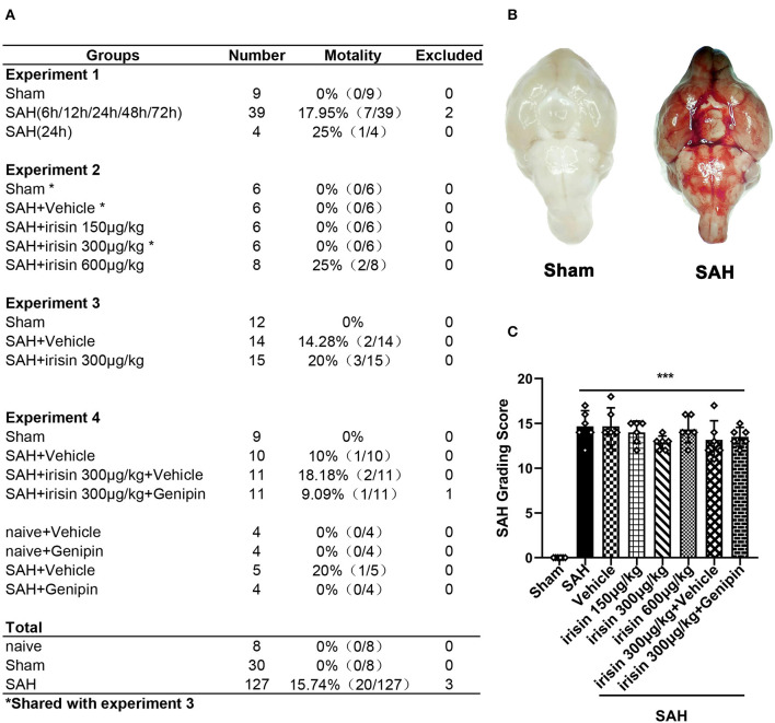
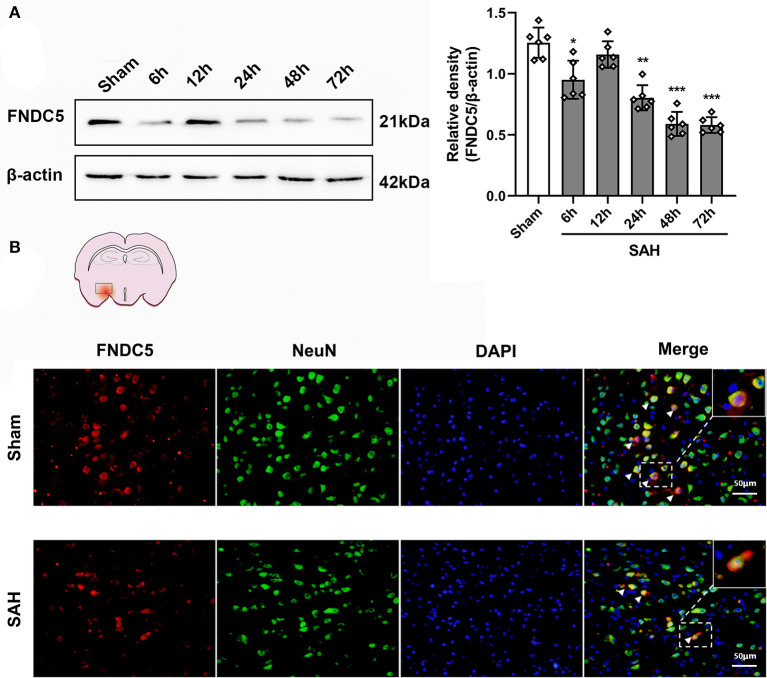
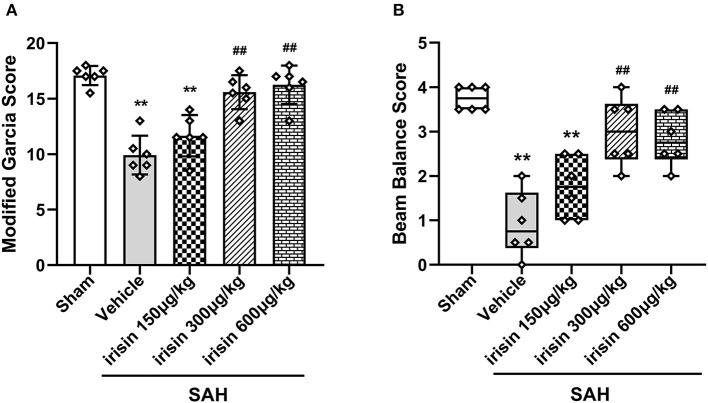
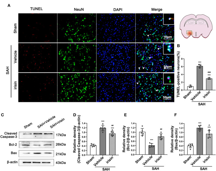
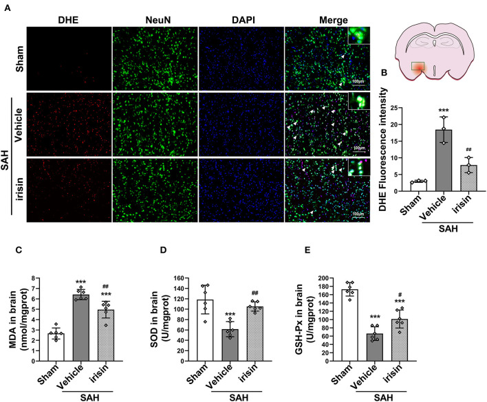
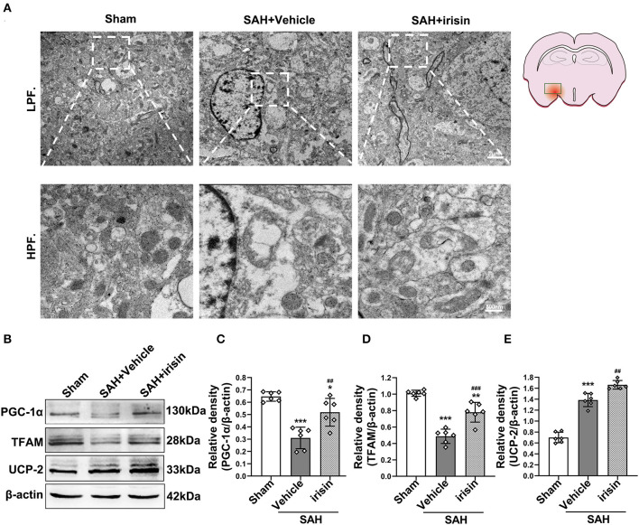
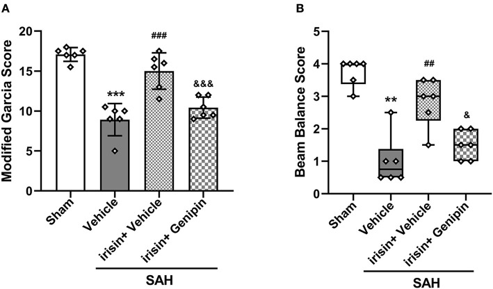
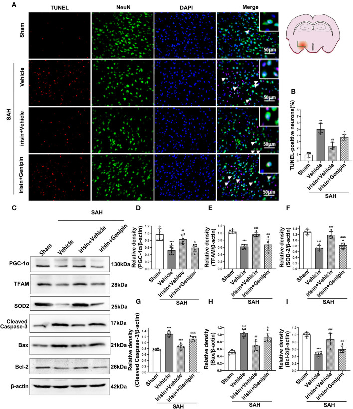
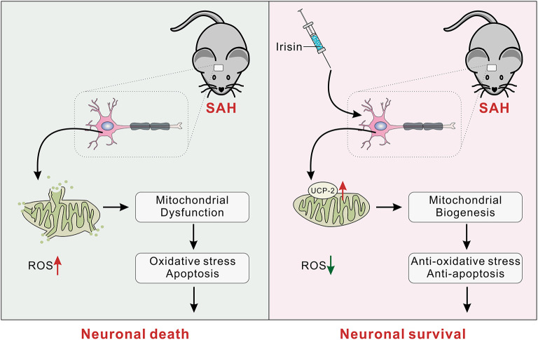
Subarachnoid hemorrhage (SAH) is a devastating form of stroke, which poses a series of intractable challenges to clinical practice. Imbalance of mitochondrial homeostasis has been thought to be the crucial pathomechanism in early brain injury (EBI) cascade after SAH. Irisin, a protein related to metabolism and mitochondrial homeostasis, has been reported to play pivotal roles in post-stroke neuroprotection. However, whether this myokine can exert neuroprotection effects after SAH remains unknown. In the present study, we explored the protective effects of irisin and the underlying mechanisms related to mitochondrial biogenesis in a SAH animal model. Endovascular perforation was used to induce SAH, and recombinant irisin was administered intracerebroventricularly. Neurobehavioral assessments, TdT-UTP nick end labeling (TUNEL) staining, dihydroethidium (DHE) staining, immunofluorescence, western blot, and transmission electron microscopy (TEM) were performed for post-SAH assessments. We demonstrated that irisin treatment improved neurobehavioral scores, reduced neuronal apoptosis, and alleviated oxidative stress in EBI after SAH. More importantly, the administration of exogenous irisin conserved the mitochondrial morphology and promoted mitochondrial biogenesis. The protective effects of irisin were partially reversed by the mitochondrial uncoupling protein-2 (UCP-2) inhibitor. Taken together, irisin may have neuroprotective effects against SAH via improving the mitochondrial biogenesis, at least in part, through UCP-2 related targets.
Keywords: FNDC5/irisin; mitochondrial homeostasis; neuronal apoptosis; oxidative stress; subarachnoid hemorrhage.
Copyright © 2021 Tu, Yin, Pang, Zhang, Zhang, Zhang, Xie, Guo, Chen, Peng and Jiang.
Conflict of interest statement
The authors declare that the research was conducted in the absence of any commercial or financial relationships that could be construed as a potential conflict of interest.
Figures
Similar articles
-
TT01001 attenuates oxidative stress and neuronal apoptosis by preventing mitoNEET-mediated mitochondrial dysfunction after subarachnoid hemorrhage in rats.Neuroreport. 2020 Aug 5;31(11):845-850. doi: 10.1097/WNR.0000000000001492. Neuroreport. 2020. PMID: 32604395
-
AVE 0991 attenuates oxidative stress and neuronal apoptosis via Mas/PKA/CREB/UCP-2 pathway after subarachnoid hemorrhage in rats.Redox Biol. 2019 Jan;20:75-86. doi: 10.1016/j.redox.2018.09.022. Epub 2018 Sep 28. Redox Biol. 2019. PMID: 30296700 Free PMC article.
-
Hydrogen-Rich Saline Attenuated Subarachnoid Hemorrhage-Induced Early Brain Injury in Rats by Suppressing Inflammatory Response: Possible Involvement of NF-κB Pathway and NLRP3 Inflammasome.Mol Neurobiol. 2016 Jul;53(5):3462-3476. doi: 10.1007/s12035-015-9242-y. Epub 2015 Jun 20. Mol Neurobiol. 2016. PMID: 26091790
-
Recombinant OX40 attenuates neuronal apoptosis through OX40-OX40L/PI3K/AKT signaling pathway following subarachnoid hemorrhage in rats.Exp Neurol. 2020 Apr;326:113179. doi: 10.1016/j.expneurol.2020.113179. Epub 2020 Jan 10. Exp Neurol. 2020. PMID: 31930990 Review.
-
Biophysical Modulation of the Mitochondrial Metabolism and Redox in Bone Homeostasis and Osteoporosis: How Biophysics Converts into Bioenergetics.Antioxidants (Basel). 2021 Aug 30;10(9):1394. doi: 10.3390/antiox10091394. Antioxidants (Basel). 2021. PMID: 34573026 Free PMC article. Review.
Cited by
-
The role of exerkines on brain mitochondria: a mini-review.J Appl Physiol (1985). 2023 Jan 1;134(1):28-35. doi: 10.1152/japplphysiol.00565.2022. Epub 2022 Nov 23. J Appl Physiol (1985). 2023. PMID: 36417200 Free PMC article. Review.
-
Inhibition of integrin alpha v/beta 5 mitigates the protective effect induced by irisin in hemorrhage.Exp Mol Pathol. 2023 Dec;134:104869. doi: 10.1016/j.yexmp.2023.104869. Epub 2023 Sep 27. Exp Mol Pathol. 2023. PMID: 37690529 Free PMC article.
-
Irisin: A Promising Target for Ischemia-Reperfusion Injury Therapy.Oxid Med Cell Longev. 2021 Oct 29;2021:5391706. doi: 10.1155/2021/5391706. eCollection 2021. Oxid Med Cell Longev. 2021. PMID: 34745418 Free PMC article. Review.
-
Preconditioning Exercise in Rats Attenuates Early Brain Injury Resulting from Subarachnoid Hemorrhage by Reducing Oxidative Stress, Inflammation, and Neuronal Apoptosis.Mol Neurobiol. 2021 Nov;58(11):5602-5617. doi: 10.1007/s12035-021-02506-7. Epub 2021 Aug 9. Mol Neurobiol. 2021. PMID: 34368932
-
The mitochondrial quality control system: a new target for exercise therapeutic intervention in the treatment of brain insulin resistance-induced neurodegeneration in obesity.Int J Obes (Lond). 2024 Jun;48(6):749-763. doi: 10.1038/s41366-024-01490-x. Epub 2024 Feb 20. Int J Obes (Lond). 2024. PMID: 38379083 Review.
References
-
- Askari H., Rajani S. F., Poorebrahim M., Haghi-Aminjan H., Raeis-Abdollahi E., Abdollahi M. (2018). A glance at the therapeutic potential of irisin against diseases involving inflammation, oxidative stress, and apoptosis: an introductory review. Pharmacol. Res. 129, 44–55. 10.1016/j.phrs.2018.01.012 - DOI - PubMed
-
- Bi J., Zhang J., Ren Y., Du Z., Li Q., Wang Y., et al. . (2019). Irisin alleviates liver ischemia-reperfusion injury by inhibiting excessive mitochondrial fission, promoting mitochondrial biogenesis and decreasing oxidative stress. Redox Biol. 20, 296–306. 10.1016/j.redox.2018.10.019 - DOI - PMC - PubMed
LinkOut - more resources
Full Text Sources
Other Literature Sources