Review
. 2021 Feb 3:11:619458.
doi: 10.3389/fphar.2020.619458. eCollection 2020.

Ectonucleotidases in Acute and Chronic Inflammation

Affiliations
Review

Ectonucleotidases in Acute and Chronic Inflammation

Anna Lisa Giuliani et al. Front Pharmacol. .

Abstract

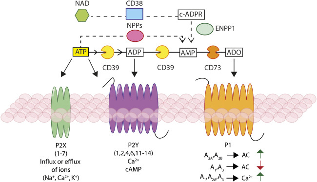
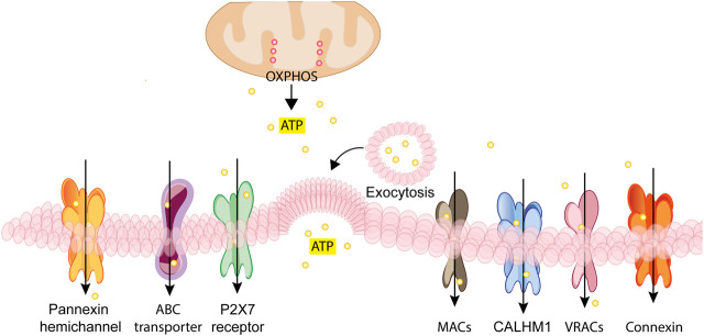
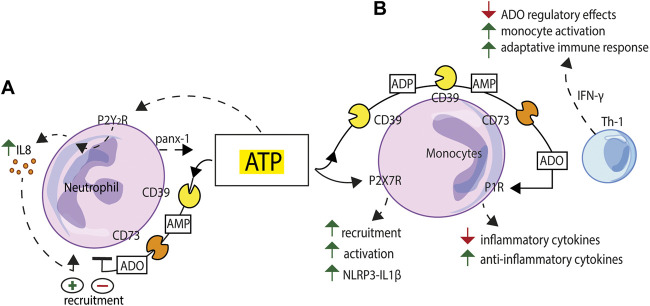
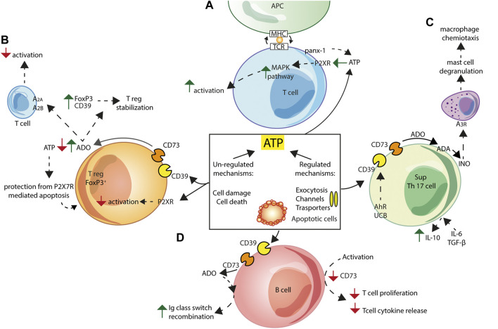
Ectonucleotidases are extracellular enzymes with a pivotal role in inflammation that hydrolyse extracellular purine and pyrimidine nucleotides, e.g., ATP, UTP, ADP, UDP, AMP and NAD+. Ectonucleotidases, expressed by virtually all cell types, immune cells included, either as plasma membrane-associated or secreted enzymes, are classified into four main families: 1) nucleoside triphosphate diphosphohydrolases (NTPDases), 2) nicotinamide adenine dinucleotide glycohydrolase (NAD glycohydrolase/ADP-ribosyl cyclase/cyclic ADP-ribose hydrolase 1), 3) ecto-5'-nucleotidase (NT5E), and 4) ecto-nucleotide pyrophosphatase/phosphodiesterases (NPPs). Concentration of ATP, UTP and NAD+ can be increased in the extracellular space thanks to un-regulated, e.g., cell damage or cell death, or regulated processes. Regulated processes include secretory exocytosis, connexin or pannexin hemichannels, ATP binding cassette (ABC) transporters, calcium homeostasis modulator (CALMH) channels, the ATP-gated P2X7 receptor, maxi-anion channels (MACs) and volume regulated ion channels (VRACs). Hydrolysis of extracellular purine nucleotides generates adenosine, an important immunosuppressant. Extracellular nucleotides and nucleosides initiate or dampen inflammation via P2 and P1 receptors, respectively. All these agents, depending on their level of expression or activation and on the agonist concentration, are potent modulators of inflammation and key promoters of host defences, immune cells activation, pathogen clearance, tissue repair and regeneration. Thus, their knowledge is of great importance for a full understanding of the pathophysiology of acute and chronic inflammatory diseases. A selection of these pathologies will be briefly discussed here.

Keywords: ATP; acute inflammation; chronic inflammatory diseases; ecto-nucleotidases; immune cells; purinergic receptors; tumors.


Conflict of interest statement

The authors declare that the research was conducted in the absence of any commercial or financial relationships that could be construed as a potential conflict of interest.

Figures

FIGURE 1
FIGURE 1
Schematic rendition of the basic elements of the purinergic signalling. Ectonucleotidases, NTPDase1/CD39, NAD glycohydrolase/CD38, NPPs and NT5E/CD73, hydrolyse extracellular ATP and NAD+, generating ADP, AMP, and adenosine (ADO). Extracellular ATP and ADP activate different P2X ionotropic and/or P2Y metabotropic receptors, leading to changes in the intracellular ion and/or cAMP concentration. Extracellular ADO stimulates P1 receptors responsible of modulation of adenylate cyclase (AC) activity and leading to changes in cAMP and Ca2+ concentration.
FIGURE 2
FIGURE 2
Schematic rendition of the different pathways for regulated nucleotide release. ATP generated inside the cell by glycolysis and oxidative phosphorylation (OXPHOS) can be released through vesicular exocytosis, connexin or pannexin channels, specific ATP binding cassette (ABC) transporters, calcium homeostasis modulators (CALHM) channels, the P2X7 receptor, maxi-anion channels (MACs) or through volume regulated ion channels (VRACs). These different pathways variably participate in ATP release in various cell types depending on the given patho-physiological context.
FIGURE 3
FIGURE 3
Schematic exemplification of purinergic receptor/ectonucleotidase cooperation in the activation/inhibition of the innate immune response. (A) ATP released via pannexin-1 (panx-1) from human neutrophils exposed to inflammatory stimuli triggers IL-8 production acting at the P2Y2R. NTPDase1/CD39 and NT5E/CD73 sequentially degrade extracellular ATP and limit neutrophil recruitment. (B) Extracellular nucleotides, firstly ATP, acting at the P2X7R, promote monocyte recruitment, modulate phagocytosis and support NLRP3 inflammasome-mediated IL-1β release. NTPDase1/CD39 and NT5E/CD73, expressed to high level on the macrophage plasma membrane, generate adenosine (ADO) and support a feed-back regulatory mechanism. Adenosine-mediated P1R stimulation generates an anti-inflammatory environment characterized by down-modulation of inflammatory cytokines release and enhanced secretion of anti-inflammatory cytokines and growth factors. This balance can be tilted towards an activated state, e.g., to support a more vigorous adaptive immune response, by IFN-γ, a stimulus that makes macrophages less sensitive to adenosine inhibition.
FIGURE 4
FIGURE 4
Schematic exemplification of purinergic receptor/ectonucleotidase cooperation in the activation/inhibition of the adaptive immune response. ATP can be released into the extracellular space via both regulated and non-regulated mechanisms. (A) In T cells, pannexin-1 (panx-1) and P2XRs localize at the immune synapse following T-cell receptor (TCR) engagement. ATP released via panx-1 triggers P2XRs activation leading to increased MAPK signalling and T cell activation. (B) Treg Foxp3+ cells generation and function are inhibited by ATP-mediated P2XRs signalling. Adenosine (ADO) formed by Treg NTPDase1/CD39 and NT5E/CD73 activity causes Treg cells stabilization, by increasing expression of Foxp3 and NTPDase1/CD39, and inhibits T cells responses acting at A2A and A2B adenosine receptors. Finally, Tregs are protected from P2X7R-mediated apoptosis thanks to extracellular ATP scavenging by NTPDase1/CD39 and NT5E/CD73. (C) Non-pathogenic Th17 cells express NTPDase1/CD39 and NT5E/CD73, and, following IL-6 and TGF-β stimulation, secrete IL-10, thus showing the typical Th17 suppressor cell (SupTh17) phenotype. NTPDase1/CD39 expression by Th17 lymphocytes is enhanced following exposure to aryl hydrocarbon receptor (AhR) agonists, such as unconjugated bilirubin (UCB). Enhanced SupTh17 adenosine deaminase (ADA) activity accelerates conversion of adenosine to inosine (INO), which activates the A3 adenosine receptor on mast cells, thus causing degranulation and release of macrophage chemotactic factors. (D) Human peripheral B cells co-express NTPDase1/CD39 and NT5E/CD73. In vitro activation of B lymphocytes co-cultured with T lymphocytes down-regulates NT5E/CD73 expression and inhibits T cell proliferation and T cell-dependent cytokine release. Extracellular adenosine contributes to immunoglobulin (Ig) class switch recombination in human naïve and IgM memory B cells.

Similar articles

Cited by

References

    1. Abascal F., Zardoya R. (2012). LRRC8 proteins share a common ancestor with pannexins, and may form hexameric channels involved in cell-cell communication. Bioessays 34 (7), 551–560. 10.1002/bies.201100173 - DOI - PubMed
    1. Abraham E. H., Prat A. G., Gerweck L., Seneveratne T., Arceci R. J., Kramer R., et al. (1993). The multidrug resistance (mdr1) gene product functions as an ATP channel. Proc. Natl. Acad. Sci. U. S. A. 90 (1), 312–316. - PMC - PubMed
    1. Adinolfi E., Capece M., Franceschini A., Falzoni S., Giuliani A. L., Rotondo A., et al. (2015). Accelerated tumor progression in mice lacking the ATP receptor P2X7. Cancer Res. 75 (4), 635–644. 10.1158/0008-5472.CAN-14-1259 - DOI - PubMed
    1. Adinolfi E., Giuliani A. L., De Marchi E., Pegoraro A., Orioli E., Di Virgilio F. (2018). The P2X7 receptor: a main player in inflammation. Biochem. Pharmacol. 151, 234–244. 10.1016/j.bcp.2017.12.021 - DOI - PubMed
    1. Adinolfi E., Raffaghello L., Giuliani A. L., Cavazzini L., Capece M., Chiozzi P., et al. (2012). Expression of P2X7 receptor increases in vivo tumor growth. Cancer Res. 72 (12), 2957–2969. 10.1158/0008-5472.CAN-11-1947 - DOI - PubMed

LinkOut - more resources