

Electrocardiographic Risk Markers of Cardiac Death: Gender Differences in the General Population
- PMID: 33613298
- PMCID: PMC7894046
- DOI: 10.3389/fphys.2020.578059
Electrocardiographic Risk Markers of Cardiac Death: Gender Differences in the General Population
Abstract
Background: Cardiac death is one of the leading causes of death and sudden cardiac death (SCD) is estimated to cause approximately 50% of cardiac deaths. Men have a higher cardiac mortality than women. Consequently, the mechanisms and risk markers of cardiac mortality are not as well defined in women as they are in men.
Aim: The aim of the study was to assess the prognostic value and possible gender differences of SCD risk markers of standard 12-lead electrocardiogram in three large general population samples.
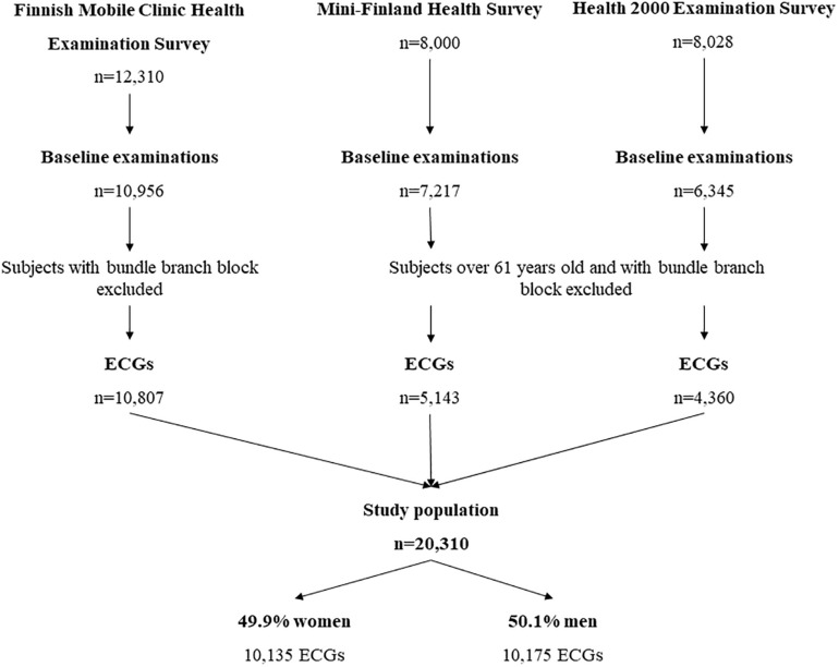
Methods: The standard 12-lead electrocardiographic (ECG) markers were analyzed from three different Finnish general population samples including total of 20,310 subjects (49.9% women, mean age 44.8 ± 8.7 years). The primary endpoint was cardiac death, and SCD and all-cause mortality were secondary endpoints. The interaction effect between women and men was assessed for each ECG variable.
Results: During the follow-up (7.7 ± 1.2 years), a total of 883 deaths occurred (24.5% women, p < 0.001). There were 296 cardiac deaths (13.9% women, p < 0.001) and 149 SCDs (14.8% women, p < 0.001). Among those who had died due to cardiac cause, women had more often a normal electrocardiogram compared to men (39.0 vs. 27.5%, p = 0.132). After adjustments with common cardiovascular risk factors and the population sample, the following ECG variables predicted the primary endpoint in men: left ventricular hypertrophy (LVH) with strain pattern (p < 0.001), QRS duration > 110 ms (p < 0.001), inferior or lateral T-wave inversion (p < 0.001) and inferolateral early repolarization (p = 0.033). In women none of the variables remained significant predictors of cardiac death in multivariable analysis, but LVH, QTc ≥ 490 ms and T-wave inversions predicted SCD (p < 0.047 and 0.033, respectively). In the interaction analysis, LVH (HR: 2.4; 95% CI: 1.2-4.9; p = 0.014) was stronger predictor of primary endpoint in women than in men.
Conclusion: Several standard ECG variables provide independent information on the risk of cardiac mortality in men but not in women. LVH and T-wave inversions predict SCD also in women.
Keywords: ECG; T wave inversion; cardiac death; gender differences; left ventricular hypertrophy; prolonged QRS; sudden cardiac death.
Copyright © 2021 Haukilahti, Kenttä, Tikkanen, Anttonen, Aro, Kerola, Eranti, Holkeri, Rissanen, Heliövaara, Knekt, Junttila and Huikuri.
Conflict of interest statement
The authors declare that the research was conducted in the absence of any commercial or financial relationships that could be construed as a potential conflict of interest.
Figures
Similar articles
-
Delayed QRS transition in the precordial leads of an electrocardiogram as a predictor of sudden cardiac death in the general population.Heart Rhythm. 2014 Dec;11(12):2254-60. doi: 10.1016/j.hrthm.2014.08.014. Epub 2014 Aug 13. Heart Rhythm. 2014. PMID: 25131180
-
ECG left ventricular hypertrophy as a risk predictor of sudden cardiac death.Int J Cardiol. 2019 Feb 1;276:125-129. doi: 10.1016/j.ijcard.2018.09.104. Epub 2018 Sep 27. Int J Cardiol. 2019. PMID: 30293667
-
Electrocardiogram as a predictor of sudden cardiac death in middle-aged subjects without a known cardiac disease.Int J Cardiol Heart Vasc. 2018 Aug 26;20:50-55. doi: 10.1016/j.ijcha.2018.08.002. eCollection 2018 Sep. Int J Cardiol Heart Vasc. 2018. PMID: 30167454 Free PMC article.
-
Regression of electrocardiographic left ventricular hypertrophy or strain is associated with lower incidence of cardiovascular morbidity and mortality in hypertensive patients independent of blood pressure reduction - A LIFE review.J Electrocardiol. 2014 Sep-Oct;47(5):630-5. doi: 10.1016/j.jelectrocard.2014.07.003. Epub 2014 Jul 3. J Electrocardiol. 2014. PMID: 25052475 Review.
-
Electrocardiographic T Wave Abnormalities and the Risk of Sudden Cardiac Death: The Finnish Perspective.Ann Noninvasive Electrocardiol. 2015 Nov;20(6):526-33. doi: 10.1111/anec.12310. Epub 2015 Sep 22. Ann Noninvasive Electrocardiol. 2015. PMID: 26391699 Free PMC article. Review.
Cited by
-
Explainable AI associates ECG aging effects with increased cardiovascular risk in a longitudinal population study.NPJ Digit Med. 2025 Jan 13;8(1):25. doi: 10.1038/s41746-024-01428-7. NPJ Digit Med. 2025. PMID: 39806125 Free PMC article.
-
Observational study of sudden cardiac arrest risk (OSCAR): Rationale and design of an electronic health records cohort.Int J Cardiol Heart Vasc. 2025 Jan 19;56:101614. doi: 10.1016/j.ijcha.2025.101614. eCollection 2025 Feb. Int J Cardiol Heart Vasc. 2025. PMID: 39897418 Free PMC article.
-
Left Ventricular Hypertrophy and Ventricular Tachyarrhythmia: The Role of Biomarkers.Int J Mol Sci. 2023 Feb 15;24(4):3881. doi: 10.3390/ijms24043881. Int J Mol Sci. 2023. PMID: 36835293 Free PMC article. Review.
-
Central Obesity is Associated with Increased Left Ventricular Maximal Wall Thickness and Intrathoracic Adipose Tissue Measured with Cardiac Magnetic Resonance.High Blood Press Cardiovasc Prev. 2024 Jul;31(4):389-399. doi: 10.1007/s40292-024-00659-9. Epub 2024 Jun 14. High Blood Press Cardiovasc Prev. 2024. PMID: 38874885 Free PMC article.
References
-
- Airaksinen J., Aalto-Setälä K., Hartikainen J., Huikuri H., Laine M., Lommi J., et al. (2016). Kardiogia: Sepelvaltimotaudin Vaaratekijät ja Ateroskleroosi, 3rd Edn Helsinki: Duodecim.
-
- Aro A. L., Anttonen O., Tikkanen J. T., Junttila M. J., Kerola T., Rissanen H. A., et al. (2011). Intraventricular conduction delay in a standard 12-lead electrocardiogram as a predictor of mortality in the general population. Circ. Arrhythm Electrophysiol. 4 704–710. 10.1161/CIRCEP.111.963561 - DOI - PubMed
-
- Aro A. L., Anttonen O., Tikkanen J. T., Junttila M. J., Kerola T., Rissanen H. A., et al. (2012). Prevalence and prognostic significance of T-Wave inversions in right precordial leads of a 12-lead electrocardiogram in the middle-aged subjects. Circulation 125 2572–2577. 10.1161/CIRCULATIONAHA.112.098681 - DOI - PubMed
LinkOut - more resources
Full Text Sources
Other Literature Sources