

Widespread Decrease of Cerebral Vimentin-Immunoreactive Astrocytes in Depressed Suicides
- PMID: 33613346
- PMCID: PMC7890082
- DOI: 10.3389/fpsyt.2021.640963
Widespread Decrease of Cerebral Vimentin-Immunoreactive Astrocytes in Depressed Suicides
Abstract
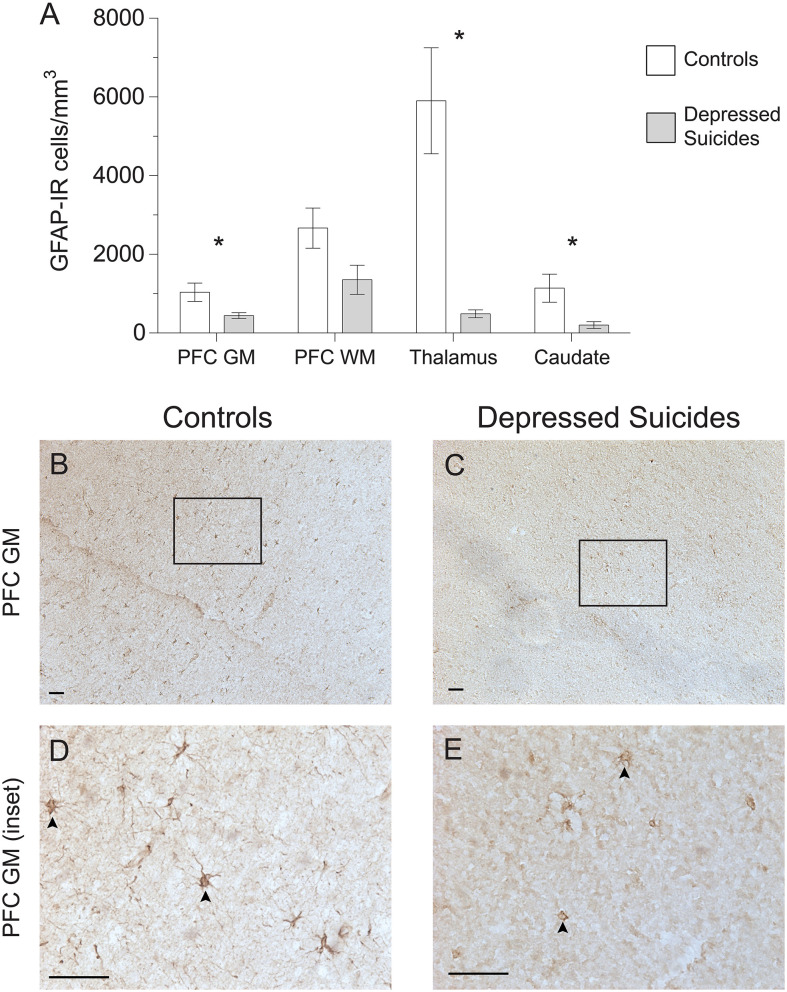
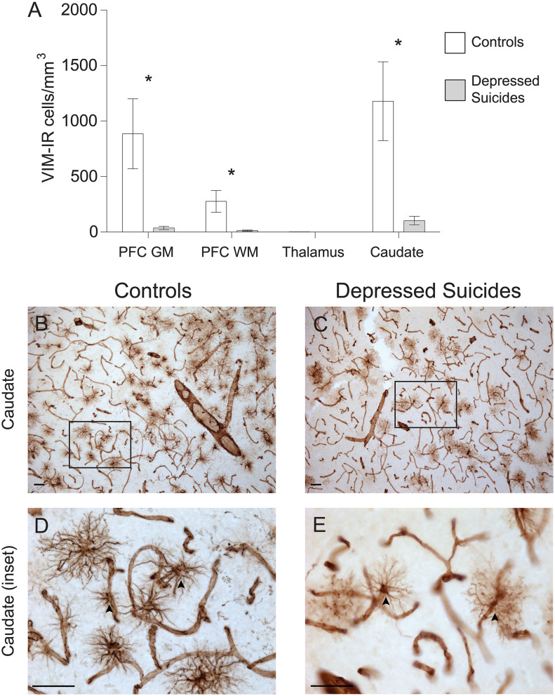
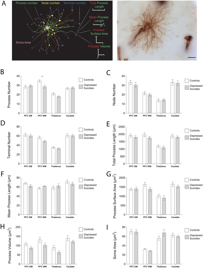
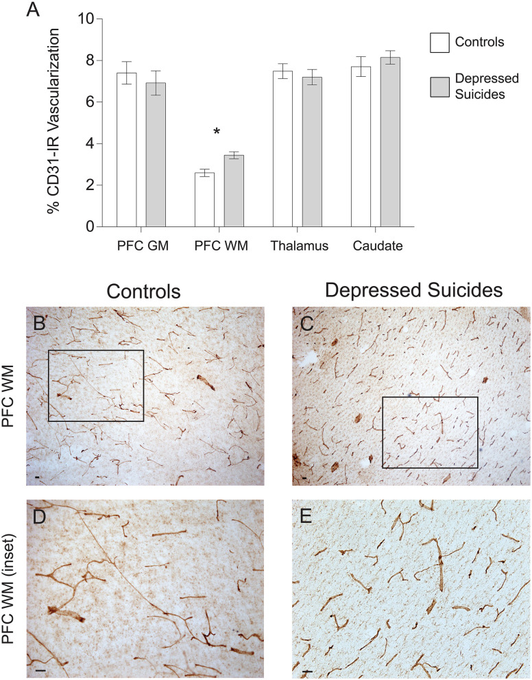
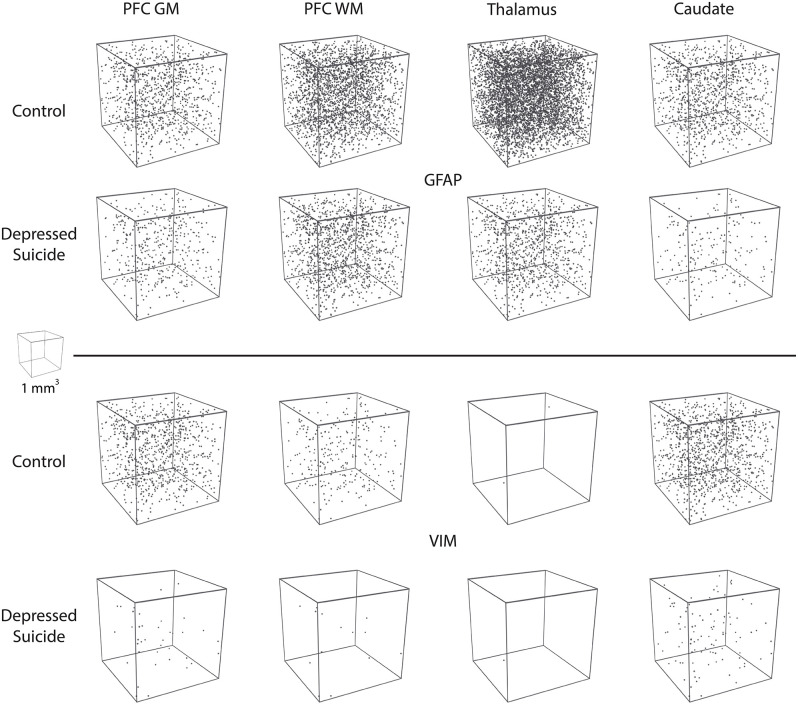
Post-mortem investigations have implicated cerebral astrocytes immunoreactive (-IR) for glial fibrillary acidic protein (GFAP) in the etiopathology of depression and suicide. However, it remains unclear whether astrocytic subpopulations IR for other astrocytic markers are similarly affected. Astrocytes IR to vimentin (VIM) display different regional densities than GFAP-IR astrocytes in the healthy brain, and so may be differently altered in depression and suicide. To investigate this, we compared the densities of GFAP-IR astrocytes and VIM-IR astrocytes in post-mortem brain samples from depressed suicides and matched non-psychiatric controls in three brain regions (dorsomedial prefrontal cortex, dorsal caudate nucleus and mediodorsal thalamus). A quantitative comparison of the fine morphology of VIM-IR astrocytes was also performed in the same regions and subjects. Finally, given the close association between astrocytes and blood vessels, we also assessed densities of CD31-IR blood vessels. Like for GFAP-IR astrocytes, VIM-IR astrocyte densities were found to be globally reduced in depressed suicides relative to controls. By contrast, CD31-IR blood vessel density and VIM-IR astrocyte morphometric features in these regions were similar between groups, except in prefrontal white matter, in which vascularization was increased and astrocytes displayed fewer primary processes. By revealing a widespread reduction of cerebral VIM-IR astrocytes in cases vs. controls, these findings further implicate astrocytic dysfunctions in depression and suicide.
Keywords: GFAP; astrocyte; depression; human; post-mortem; suicide; vimentin.
Copyright © 2021 O'Leary, Belliveau, Davoli, Ma, Tanti, Turecki and Mechawar.
Conflict of interest statement
The authors declare that the research was conducted in the absence of any commercial or financial relationships that could be construed as a potential conflict of interest.
Figures
Similar articles
-
Characterization of Vimentin-Immunoreactive Astrocytes in the Human Brain.Front Neuroanat. 2020 Jul 30;14:31. doi: 10.3389/fnana.2020.00031. eCollection 2020. Front Neuroanat. 2020. PMID: 32848635 Free PMC article.
-
Glial fibrillary acidic protein is differentially expressed across cortical and subcortical regions in healthy brains and downregulated in the thalamus and caudate nucleus of depressed suicides.Mol Psychiatry. 2016 Apr;21(4):509-15. doi: 10.1038/mp.2015.65. Epub 2015 Jun 2. Mol Psychiatry. 2016. PMID: 26033239
-
Implication of cerebral astrocytes in major depression: A review of fine neuroanatomical evidence in humans.Glia. 2021 Sep;69(9):2077-2099. doi: 10.1002/glia.23994. Epub 2021 Mar 18. Glia. 2021. PMID: 33734498 Review.
-
Evidence for increased microglial priming and macrophage recruitment in the dorsal anterior cingulate white matter of depressed suicides.Brain Behav Immun. 2014 Nov;42:50-9. doi: 10.1016/j.bbi.2014.05.007. Epub 2014 May 20. Brain Behav Immun. 2014. PMID: 24858659
-
Astrocytes in the Neuropathology of Bipolar Disorder: Review of Current Evidence.Brain Sci. 2022 Nov 8;12(11):1513. doi: 10.3390/brainsci12111513. Brain Sci. 2022. PMID: 36358439 Free PMC article. Review.
Cited by
-
Whole-exome sequencing identifies protein-coding variants associated with brain iron in 29,828 individuals.Nat Commun. 2024 Jul 2;15(1):5540. doi: 10.1038/s41467-024-49702-2. Nat Commun. 2024. PMID: 38956042 Free PMC article.
-
Astrocytes in Post-Stroke Depression: Roles in Inflammation, Neurotransmission, and Neurotrophin Signaling.Cell Mol Neurobiol. 2023 Oct;43(7):3301-3313. doi: 10.1007/s10571-023-01386-w. Epub 2023 Jul 20. Cell Mol Neurobiol. 2023. PMID: 37470888 Free PMC article. Review.
-
Unraveling the Role of the Blood-Brain Barrier in the Pathophysiology of Depression: Recent Advances and Future Perspectives.Mol Neurobiol. 2024 Dec;61(12):10398-10447. doi: 10.1007/s12035-024-04205-5. Epub 2024 May 10. Mol Neurobiol. 2024. PMID: 38730081 Review.
-
Exploring neurometabolic alterations in bipolar disorder with suicidal ideation based on proton magnetic resonance spectroscopy and machine learning technology.Front Neurosci. 2022 Sep 9;16:944585. doi: 10.3389/fnins.2022.944585. eCollection 2022. Front Neurosci. 2022. PMID: 36161155 Free PMC article.
-
Astrocytes in human central nervous system diseases: a frontier for new therapies.Signal Transduct Target Ther. 2023 Oct 13;8(1):396. doi: 10.1038/s41392-023-01628-9. Signal Transduct Target Ther. 2023. PMID: 37828019 Free PMC article. Review.
References
LinkOut - more resources
Full Text Sources
Other Literature Sources
Miscellaneous