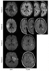
Extent of FLAIR Hyperintense Vessels May Modify Treatment Effect of Thrombolysis: A Post hoc Analysis of the WAKE-UP Trial
- PMID: 33613422
- PMCID: PMC7890254
- DOI: 10.3389/fneur.2020.623881
Extent of FLAIR Hyperintense Vessels May Modify Treatment Effect of Thrombolysis: A Post hoc Analysis of the WAKE-UP Trial
Abstract
Background and Aims: Fluid-attenuated inversion recovery (FLAIR) hyperintense vessels (FHVs) on MRI are a radiological marker of vessel occlusion and indirect sign of collateral circulation. However, the clinical relevance is uncertain. We explored whether the extent of FHVs is associated with outcome and how FHVs modify treatment effect of thrombolysis in a subgroup of patients with confirmed unilateral vessel occlusion from the randomized controlled WAKE-UP trial. Methods: One hundred sixty-five patients were analyzed. Two blinded raters independently assessed the presence and extent of FHVs (defined as the number of slices with visible FHV multiplied by FLAIR slice thickness). Patients were then separated into two groups to distinguish between few and extensive FHVs (dichotomization at the median <30 or ≥30). Results: Here, 85% of all patients (n = 140) and 95% of middle cerebral artery (MCA) occlusion patients (n = 127) showed FHVs at baseline. Between MCA occlusion patients with few and extensive FHVs, no differences were identified in relative lesion growth (p = 0.971) and short-term [follow-up National Institutes of Health Stroke Scale (NIHSS) score; p = 0.342] or long-term functional recovery [modified Rankin Scale (mRS) <2 at 90 days poststroke; p = 0.607]. In linear regression analysis, baseline extent of FHV (defined as a continuous variable) was highly associated with volume of hypoperfused tissue (β = 2.161; 95% CI 0.96-3.36; p = 0.001). In multivariable regression analysis adjusted for treatment group, stroke severity, lesion volume, occlusion site, and recanalization, FHV did not modify functional recovery. However, in patients with few FHVs, the odds for good functional outcome (mRS) were increased in recombinant tissue plasminogen activator (rtPA) patients compared to those who received placebo [odds ratio (OR) = 5.3; 95% CI 1.2-24.0], whereas no apparent benefit was observed in patients with extensive FHVs (OR = 1.1; 95% CI 0.3-3.8), p-value for interaction was 0.11. Conclusion: While the extent of FHVs on baseline did not alter the evolution of stroke in terms of lesion progression or functional recovery, it may modify treatment effect and should therefore be considered relevant additional information in those patients who are eligible for intravenous thrombolysis. Clinical Trial Registration: Main trial (WAKE-UP): ClinicalTrials.gov, NCT01525290; and EudraCT, 2011-005906-32. Registered February 2, 2012.
Keywords: FLAIR hyperintensities; MRI; hyperintense vessel; ischemic stroke; prognosis; thrombolysis; wake-up stroke.
Copyright © 2021 Grosch, Kufner, Boutitie, Cheng, Ebinger, Endres, Fiebach, Fiehler, Königsberg, Lemmens, Muir, Nighoghossian, Pedraza, Siemonsen, Thijs, Wouters, Gerloff, Thomalla and Galinovic.
Conflict of interest statement
JBF reports consulting and advisory board fees from BioClinica, Cerevast, Abbvie, AC Immune, Artemida, Brainomix, Biogen, BMS, Daiichi-Sankyo, Guerbet, Ionis Pharmaceuticals, Julius Clinical, Eli Lilly, Tau Rx, and EISAI outside the submitted work. MEn reports grants from Bayer and fees paid to the Charité from Bayer, Boehringer Ingelheim, BMS, Daiichi Sankyo, Amgen, GSK, Sanofi, Covidien, Novartis, and Pfizer, all outside the submitted work. The remaining authors declare that the research was conducted in the absence of any commercial or financial relationships that could be construed as a potential conflict of interest.
Figures


Similar articles
-
Current Smoking Does Not Modify the Treatment Effect of Intravenous Thrombolysis in Acute Ischemic Stroke Patients-A Post-hoc Analysis of the WAKE-UP Trial.Front Neurol. 2019 Nov 22;10:1239. doi: 10.3389/fneur.2019.01239. eCollection 2019. Front Neurol. 2019. PMID: 31824412 Free PMC article.
-
Fluid-attenuated inversion recovery hyperintense vessels in posterior cerebral artery infarction.Cerebrovasc Dis Extra. 2013 Apr 5;3(1):46-54. doi: 10.1159/000350459. eCollection 2013. Cerebrovasc Dis Extra. 2013. PMID: 24052794 Free PMC article.
-
Quantitative Signal Intensity in Fluid-Attenuated Inversion Recovery and Treatment Effect in the WAKE-UP Trial.Stroke. 2020 Jan;51(1):209-215. doi: 10.1161/STROKEAHA.119.027390. Epub 2019 Oct 30. Stroke. 2020. PMID: 31662118 Clinical Trial.
-
Validation of FLAIR hyperintense lesions as imaging biomarkers to predict the outcome of acute stroke after intra-arterial thrombolysis following intravenous tissue plasminogen activator.Cerebrovasc Dis. 2013;35(5):461-8. doi: 10.1159/000350201. Epub 2013 May 31. Cerebrovasc Dis. 2013. PMID: 23735898
-
Extending thrombolysis to 4·5-9 h and wake-up stroke using perfusion imaging: a systematic review and meta-analysis of individual patient data.Lancet. 2019 Jul 13;394(10193):139-147. doi: 10.1016/S0140-6736(19)31053-0. Epub 2019 May 22. Lancet. 2019. PMID: 31128925
Cited by
-
Fluid-Attenuated Inversion Recovery Vascular Hyperintensity in Cerebrovascular Disease: A Review for Radiologists and Clinicians.Front Aging Neurosci. 2021 Dec 16;13:790626. doi: 10.3389/fnagi.2021.790626. eCollection 2021. Front Aging Neurosci. 2021. PMID: 34975459 Free PMC article. Review.
-
Prognostic value of post-treatment fluid-attenuated inversion recovery vascular hyperintensity in ischemic stroke after endovascular thrombectomy.Eur Radiol. 2022 Dec;32(12):8067-8076. doi: 10.1007/s00330-022-08886-1. Epub 2022 Jun 3. Eur Radiol. 2022. PMID: 35665844
-
Editorial comment: Prognostic value of post-treatment fluid-attenuated inversion recovery vascular hyperintensity in ischemic stroke after endovascular thrombectomy.Eur Radiol. 2022 Dec;32(12):8065-8066. doi: 10.1007/s00330-022-09099-2. Epub 2022 Sep 8. Eur Radiol. 2022. PMID: 36074265
-
Stability of MRI radiomic features according to various imaging parameters in fast scanned T2-FLAIR for acute ischemic stroke patients.Sci Rep. 2021 Aug 25;11(1):17143. doi: 10.1038/s41598-021-96621-z. Sci Rep. 2021. PMID: 34433881 Free PMC article.
-
Estimating Perfusion Deficits in Acute Stroke Patients Without Perfusion Imaging.Stroke. 2022 Nov;53(11):3439-3445. doi: 10.1161/STROKEAHA.121.038101. Epub 2022 Jul 22. Stroke. 2022. PMID: 35866426 Free PMC article.
References
Associated data
LinkOut - more resources
Full Text Sources
Other Literature Sources
Medical

