Review
. 2021 Feb 4:12:612675.
doi: 10.3389/fmicb.2021.612675. eCollection 2021.

Evolution of Drug-Resistant Mycobacterium tuberculosis Strains and Their Adaptation to the Human Lung Environment

Affiliations
Review

Evolution of Drug-Resistant Mycobacterium tuberculosis Strains and Their Adaptation to the Human Lung Environment

Anna Allué-Guardia et al. Front Microbiol. .

Abstract

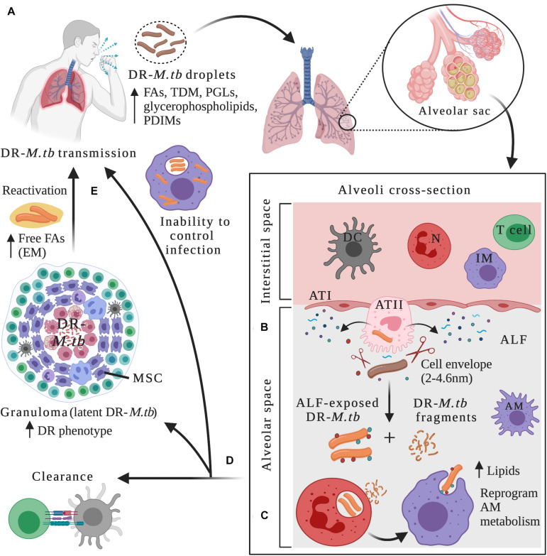
In the last two decades, multi (MDR), extensively (XDR), extremely (XXDR) and total (TDR) drug-resistant Mycobacterium tuberculosis (M.tb) strains have emerged as a threat to public health worldwide, stressing the need to develop new tuberculosis (TB) prevention and treatment strategies. It is estimated that in the next 35 years, drug-resistant TB will kill around 75 million people and cost the global economy $16.7 trillion. Indeed, the COVID-19 pandemic alone may contribute with the development of 6.3 million new TB cases due to lack of resources and enforced confinement in TB endemic areas. Evolution of drug-resistant M.tb depends on numerous factors, such as bacterial fitness, strain's genetic background and its capacity to adapt to the surrounding environment, as well as host-specific and environmental factors. Whole-genome transcriptomics and genome-wide association studies in recent years have shed some insights into the complexity of M.tb drug resistance and have provided a better understanding of its underlying molecular mechanisms. In this review, we will discuss M.tb phenotypic and genotypic changes driving resistance, including changes in cell envelope components, as well as recently described intrinsic and extrinsic factors promoting resistance emergence and transmission. We will further explore how drug-resistant M.tb adapts differently than drug-susceptible strains to the lung environment at the cellular level, modulating M.tb-host interactions and disease outcome, and novel next generation sequencing (NGS) strategies to study drug-resistant TB.

Keywords: Mycobacterium tuberculosis; bacterial–host interactions; drug resistance; evolution; next generation sequencing.


Conflict of interest statement

The authors declare that the research was conducted in the absence of any commercial or financial relationships that could be construed as a potential conflict of interest.

Figures

FIGURE 1
FIGURE 1
Drug-resistant M.tb–host interactions within lung microenvironments at different stages of the infection. (A) After being in close contact with an individual with active TB disease, infection can be initiated through inhalation of drug-resistant (DR) M.tb-containing droplets. DR-M.tb bacilli contain altered levels of cell envelope lipids such as free fatty acids (FAs), trehalose dimycolate (TDM), Phthiocerol dimycocerosates (PDIMs), phenolic glycolipids (PGLs) and glycerophospholipids, among others. Upon bypassing upper respiratory tract barriers, DR-M.tb will ultimately reach the alveoli, a sac-like structure composed of a thin layer of alveolar epithelial cells type I (ATIs, with structural and gas exchange function) and alveolar epithelial cells type II (ATII, with secretor function) surrounded by capillaries. Alveolar macrophages (AMs) are resident phagocytes that populate the alveolar space, while the interstitial space surrounding the alveoli contains interstitial macrophages (IMs), dendritic cells (DCs), neutrophils (N), and T cells, among other host cells. (B) In the alveolar space, DR-M.tb bacilli first interact with host soluble innate components present in the alveolar lining fluid (ALF), where hydrolases (represented as scissors) can cleave and modify the M.tb cell envelope, releasing cell envelope fragments into the alveolar space. (C) Subsequently, ALF-modified DR-M.tb bacilli will interact with AMs (professional phagocytes) and/or with ATs (non-professional phagocytes), as well as with other host innate immune cells (e.g., neutrophils, DCs). Released DR-M.tb fragments are immunogenic and could attract neutrophils to the infection site driving local oxidative stress and inflammation, which could assist resident resting AMs to clear the infection. (D) The outcome of these initial interactions will resolve in ALF-exposed DR-M.tb clearance, establishment of a successful infection driving primary active TB disease, or a latent M.tb infection defined by M.tb persisters within granulomas, a niche that provide a protective environment against anti-TB drugs, thus increasing the DR-M.tb phenotype. Surrounding mesenchymal stem cells (MSCs) can also dampen immune responses and provide a protective intracellular environment for M.tb persistence. (E) Reactivation and subsequent progression to active TB disease can happen when granulomas fail to contain DR-M.tb, with extracellular DR-M.tb growth that leads to lung tissue destruction and cavity formation. It has been suggested that in this scenario, DR-M.tb secretes free fatty acids, creating some kind of extracellular matrix (EM) that further shields the DR-M.tb against TB drugs. Figure created with BioRender.com.

Similar articles

Cited by

References

    1. Adams K. N., Takaki K., Connolly L. E., Wiedenhoft H., Winglee K., Humbert O., et al. (2011). Drug tolerance in replicating mycobacteria mediated by a macrophage-induced efflux mechanism. Cell 145 39–53. 10.1016/j.cell.2011.02.022 - DOI - PMC - PubMed
    1. Al-Saeedi M., Al-Hajoj S. (2017). Diversity and evolution of drug resistance mechanisms in Mycobacterium tuberculosis. Infect. Drug Resist. 10 333–342. 10.2147/IDR.S144446 - DOI - PMC - PubMed
    1. Arcos J., Diangelo L. E., Scordo J. M., Sasindran S. J., Moliva J. I., Turner J., et al. (2015). Lung mucosa lining fluid modification of Mycobacterium tuberculosis to reprogram human neutrophil killing mechanisms. J. Infect. Dis. 212 948–958. 10.1093/infdis/jiv146 - DOI - PMC - PubMed
    1. Arcos J., Sasindran S. J., Fujiwara N., Turner J., Schlesinger L. S., Torrelles J. B. (2011). Human lung hydrolases delineate Mycobacterium tuberculosis-macrophage interactions and the capacity to control infection. J. Immunol. 187 372–381. 10.4049/jimmunol.1100823 - DOI - PMC - PubMed
    1. Arcos J., Sasindran S. J., Moliva J. I., Scordo J. M., Sidiki S., Guo H., et al. (2017). Mycobacterium tuberculosis cell wall released fragments by the action of the human lung mucosa modulate macrophages to control infection in an IL-10-dependent manner. Mucosal Immunol. 10 1248–1258. 10.1038/mi.2016.115 - DOI - PMC - PubMed

LinkOut - more resources