Review
. 2021 Feb 3:11:609161.
doi: 10.3389/fimmu.2020.609161. eCollection 2020.

Abdominal Aortic Aneurysm: Roles of Inflammatory Cells

Affiliations
Review

Abdominal Aortic Aneurysm: Roles of Inflammatory Cells

Zhen Yuan et al. Front Immunol. .

Abstract

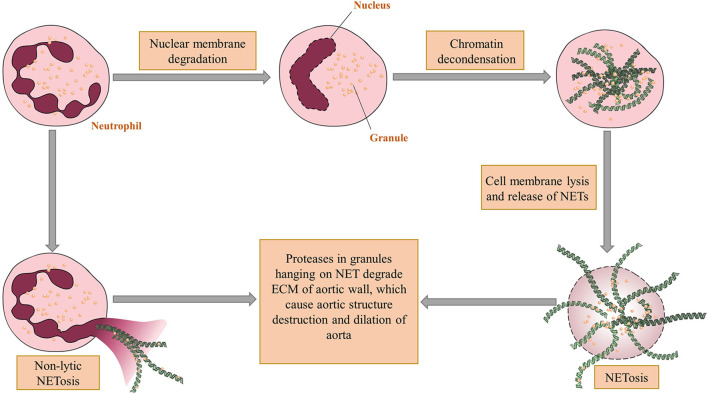
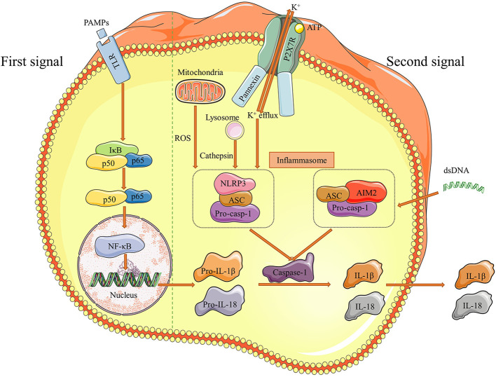
Abdominal aortic aneurysms (AAAs) are local dilations of infrarenal segment of aortas. Molecular mechanisms underlying the pathogenesis of AAA remain not fully clear. However, inflammation has been considered as a central player in the development of AAA. In the past few decades, studies demonstrated a host of inflammatory cells, including T cells, macrophages, dendritic cells, neutrophils, B cells, and mast cells, etc. infiltrating into aortic walls, which implicated their crucial roles. In addition to direct cell contacts and cytokine or protease secretions, special structures like inflammasomes and neutrophil extracellular traps have been investigated to explore their functions in aneurysm formation. The above-mentioned inflammatory cells and associated structures may initiate and promote AAA expansion. Understanding their impacts and interaction networks formation is meaningful to develop new strategies of screening and pharmacological interventions for AAA. In this review, we aim to discuss the roles and mechanisms of these inflammatory cells in AAA pathogenesis.

Keywords: T cells; abdominal aortic aneurysm; inflammasome; inflammation; macrophages; neutrophil extracellular traps.


Conflict of interest statement

The authors declare that the research was conducted in the absence of any commercial or financial relationships that could be construed as a potential conflict of interest.

Figures

Figure 1
Figure 1
The mechanism of NET formation and acting on aortic walls. There are two ways for neutrophil extracellular traps (NET) come into being. The first one is called NETosis in which nuclei of neutrophils undergo delobulation, chromatin decondensation and nuclear membrane lysis. After that neutrophil granules adhering to released chromatin enter extracellular spaces through ruptured cell membranes. The other way, which is a non-lytic form of NETosis, occurs after partial depolarization of nuclei and render granules hanging on chromatin out of plasma without cell deaths. The proteases within granules can thereby directly degrade the vascular structure and cause aortic dilation. Figures were produced using Servier Medical Art (www.servier.com).
Figure 2
Figure 2
Interactions of between B cells and other immune cells in AAA. B cells can differentiate into plasma cells and memory B cells under the stimulation of IL-4 from Th cells. Plasma cells continuously secrete immunoglobulins, which directly attack aortic walls. Specifically, IgE can activate macrophage polarization and mast cell degranulation and subsequently increase their productions of proteases such as MMPs and cathepsins. These factors work together in the pathogenesis of extracellular matrix degradation of aorta, and is an example of immune cell interactions in the whole process of AAA development. Figures were produced using Servier Medical Art (www.servier.com).
Figure 3
Figure 3
Pathways of NLRP3 and AIM2 inflammasome activation. There are two distinct signals needed for inflammasome to be effective. Initially, pathogen-associated molecular patterns (PAMPs) as the first signal binds to Toll like receptors (TLRs) and stimulate NF-κB, which increases downstream pro-IL-1β and pro-IL-18 production. Then, efflux of K+ and dsDNA are the second signals correspondingly to induce NLRP3 and AIM2 inflammasome formation. The pathway of NLRP3 inflammasome activation usually proceed under the assistant of cathepsin released by lysosome and ROS mtDNA from mitochondria. The final result of inflammasome activation is cleaving pro-casp-1 into caspase-1, which transforms pro-IL-1β and pro-IL-18 to IL-1β and IL-18. These two effective cytokines are secreted out and participate the inflammatory responses in aortic walls. Figures were produced using Servier Medical Art (www.servier.com).

References

    1. Klink A, Hyafil F, Rudd J, Faries P, Fuster V, Mallat Z, et al. Diagnostic and therapeutic strategies for small abdominal aortic aneurysms. Nat Rev Cardiol (2011) 8(6):338–47. 10.1038/nrcardio.2011.1 - DOI - PubMed
    1. Umebayashi R, Uchida HA, Wada J. Abdominal aortic aneurysm in aged population. Aging (Albany NY) (2018) 10(12):3650–1. 10.18632/aging.101702 - DOI - PMC - PubMed
    1. Lindquist Liljeqvist M, Hultgren R, Bergman O, Villard C, Kronqvist M, Eriksson P, et al. Tunica-Specific Transcriptome of Abdominal Aortic Aneurysm and the Effect of Intraluminal Thrombus, Smoking, and Diameter Growth Rate. Arterioscler Thromb Vasc Biol (2020) 40(11):2700–13. 10.1161/ATVBAHA.120.314264 - DOI - PubMed
    1. Arnaoutakis DJ, Upchurch GR., Jr. Abdominal Aortic Aneurysm Screening Is Safe yet Lacks Effectiveness. Circulation (2019) 139(11):1381–3. 10.1161/CIRCULATIONAHA.118.038809 - DOI - PubMed
    1. Eckstein HH, Bockler D, Flessenkamper I, Schmitz-Rixen T, Debus S, Lang W. Ultrasonographic screening for the detection of abdominal aortic aneurysms. Dtsch Arztebl Int (2009) 106(41):657–63. 10.3238/arztebl.2009.0657 - DOI - PMC - PubMed

Publication types

MeSH terms