. 2021 Feb 4:11:622162.
doi: 10.3389/fimmu.2020.622162. eCollection 2020.

Vitamin B6 Acquisition and Metabolism in Schistosoma mansoni

Affiliations

Vitamin B6 Acquisition and Metabolism in Schistosoma mansoni

Akram A Da'dara et al. Front Immunol. .

Abstract

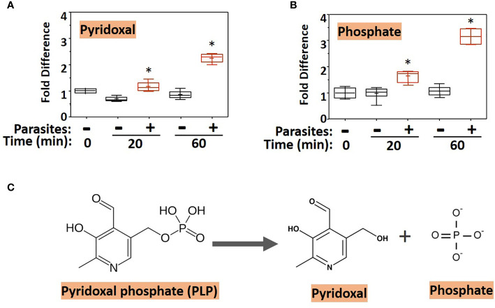
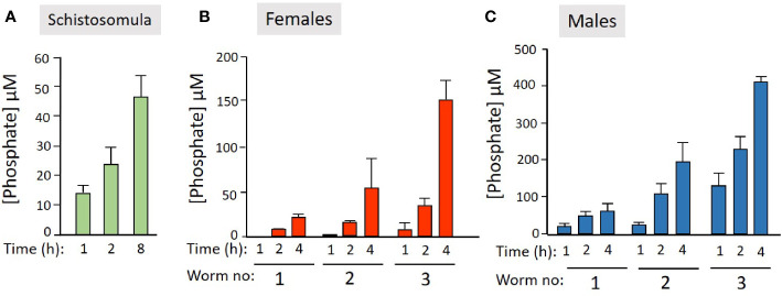
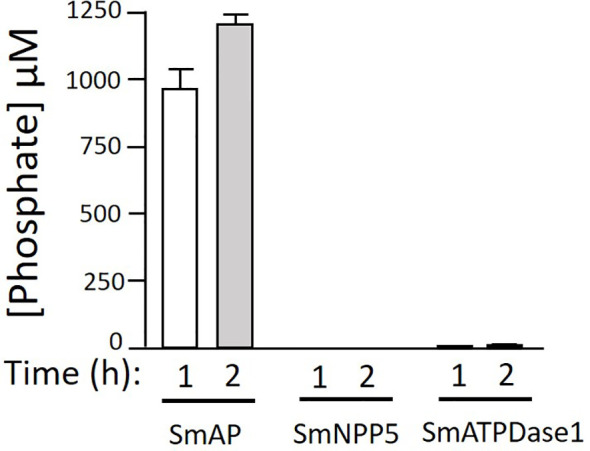
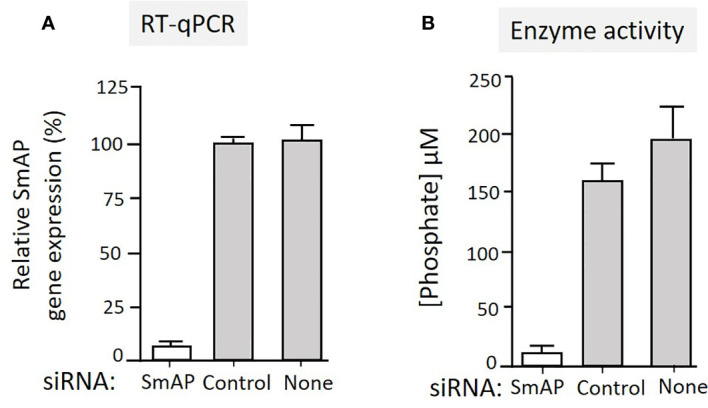
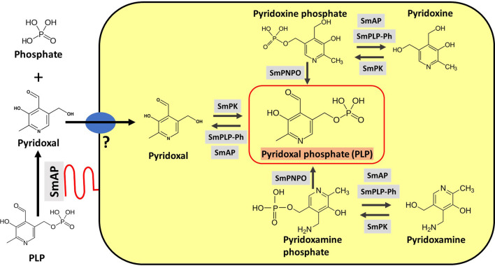
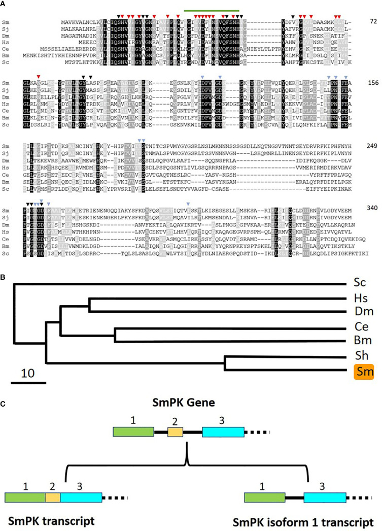
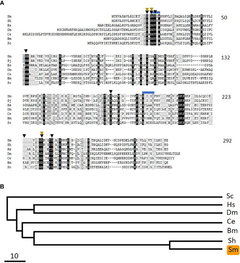
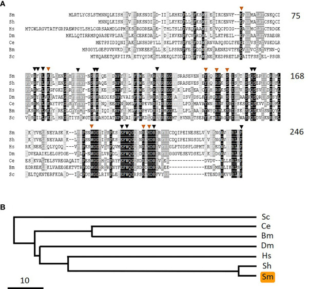
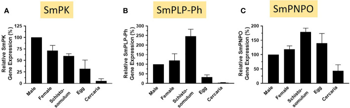
Schistosomes are parasitic platyhelminths that currently infect >200 million people globally. The adult worms can live within the vasculature of their hosts for many years where they acquire all nutrients necessary for their survival and growth. In this work we focus on how Schistosoma mansoni parasites acquire and metabolize vitamin B6, whose active form is pyridoxal phosphate (PLP). We show here that live intravascular stage parasites (schistosomula and adult males and females) can cleave exogenous PLP to liberate pyridoxal. Of the three characterized nucleotide-metabolizing ectoenzymes expressed at the schistosome surface (SmAP, SmNPP5, and SmATPDase1), only SmAP hydrolyzes PLP. Heat-inactivated recombinant SmAP can no longer cleave PLP. Further, parasites whose SmAP gene has been suppressed by RNAi are significantly impaired in their ability to cleave PLP compared to controls. When schistosomes are incubated in murine plasma, they alter its metabolomic profile-the levels of both pyridoxal and phosphate increase over time, a finding consistent with the action of host-exposed SmAP acting on PLP. We hypothesize that SmAP-mediated dephosphorylation of PLP generates a pool of pyridoxal around the worms that can be conveniently taken in by the parasites to participate in essential, vitamin B6-driven metabolism. In addition, since host PLP-dependent enzymes play active roles in inflammatory processes, parasite-mediated cleavage of this metabolite may serve to limit parasite-damaging inflammation. In this work we also identified schistosome homologs of enzymes that are involved in intracellular vitamin B6 metabolism. These are pyridoxal kinase (SmPK) as well as pyridoxal phosphate phosphatase (SmPLP-Ph) and pyridox(am)ine 5'-phosphate oxidase (SmPNPO) and cDNAs encoding these three enzymes were cloned and sequenced. The three genes encoding these enzymes all display high relative expression in schistosomula and adult worms suggestive of robust vitamin B6 metabolism in the intravascular life stages.

Keywords: PLP; alkaline phosphatase; ectoenzyme; parasite; pyridoxal phosphate; schistosome.


Conflict of interest statement

The authors declare that the research was conducted in the absence of any commercial or financial relationships that could be construed as a potential conflict of interest.

Figures

Figure 1
Figure 1
Box plots showing relative levels of pyridoxal (A) and phosphate (B) in murine plasma that either contained adult schistosomes (+, red) or did not contain schistosomes (-, black) for the indicated time periods. * indicates statistically significant difference compared to the same time point lacking parasites; Welch’s two-sample t-test, P < 0.05. Each box bounds the upper and lower quartile, the line in each box is the median value and “+” signifies the mean value for the sample; error bars indicate the maximum (upper) and minimum (lower) distribution. Values obtained at zero time (0) are set at 1. (C) Chemical structure of pyridoxal phosphate (PLP, left) and the products of its hydrolysis (indicated by the arrow), pyridoxal and phosphate (right).
Figure 2
Figure 2
Live schistosome parasites hydrolyze PLP. Phosphate generation (µM, mean +/-SD) by live schistosomula (groups of 2,000, green bars, n = 4, A) or adult females (red bars, n = 5, B) or adult males (blue bars, n = 5, C) in the presence of pyridoxal phosphate (PLP) at the indicated time points. Adult worms were either incubated singly or in groups of two or three, as indicated.
Figure 3
Figure 3
rSmAP can hydrolyze PLP. Phosphate generation (µM, mean +/-SD) by recombinant SmAP, SmNPP5, or SmATPDase1 in assay buffer containing PLP as substrate, at the indicated time points. No appreciable levels of phosphate were detected when rSmNPP5 or rSmATPDase1 were tested.
Figure 4
Figure 4
(A) Relative SmAP gene expression determined by RT-qPCR in adult male worms 48 h after treatment with an siRNA targeting the SmAP gene or with an irrelevant siRNA (Control) or with no siRNA (None), as indicated. The expression level displayed by the control worms is set at 100%. (B) Phosphate generation (µM, mean +/-SD, n = 5) by individual live adult male worms seven days after treatment with SmAP siRNA or an irrelevant siRNA (Control) or no siRNA (None) in the presence of PLP. Parasites treated with SmAP siRNA generate significantly less phosphate compared to either control (One-way ANOVA, P < 0.001).
Figure 5
Figure 5
Vitamin B6 metabolism in schistosomes. SmAP (depicted at lower left, red) can cleave PLP to generate free pyridoxal which may be imported by schistosomes via an unidentified transporter protein (blue oval, left). Depicted in the yellow rectangle are hypothetical biochemical pathways in S. mansoni leading to the generation of PLP (bounded by the red box, center). Imported pyridoxal (left) may be phosphorylated by S. mansoni pyridoxal kinase (SmPK) to directly generate PLP; the vitamers pyridoxine (top) and pyridoxamine (bottom) may first be phosphorylated by SmPK to generate pyridoxine phosphate and pyridoxamine phosphate, respectively and these products may then be acted upon by S. mansoni pyridox(am)ine-5’-phosphate oxidase (SmPNPO) to generate PLP. Dephosphorylation of several vitamers may be driven by S. mansoni pyridoxal phosphate phosphatase (SmPLP-Ph) and/or SmAP.
Figure 6
Figure 6
Alignment of SmPK with other members of the pyridoxal kinase protein family. Sm, Schistosoma mansoni (GeneBank accession number MW148599); Sj, Schistosoma japonicum (CAX69335.1); Hs, Homo sapiens (NP_003672); Dm, Drosophila melanogaster (AAR82765); Ce, Caenorhabditis elegans (NP_491463.2); Bm, Brugia malayi (VI094344); Sc, Saccharomyces cerevisiae (NP_014424). Residues found in all seven sequences are depicted as white on a black background; those common to six of the seven pyridoxal kinase homologs are white on a dark grey background and those found in a majority of the sequences (four or five out of seven) are white on a light grey background. Residues predicted to be involved in pyridoxal binding are indicated by black arrowheads; those involved in ATP binding by blue arrowheads and those involved in dimer formation, red arrowheads. The green line (top) indicates the sequence motif 36LGIEVDFINSVQFSNH51 that is replaced by 36CVLLIITY43 in the SmPK isoform 1 sequence reported in this work (GeneBank accession number of SmPK isoform 1: MW148600). Numbers at right are an amino acid count for the S. mansoni protein. (B) Phylogenetic tree (absolute distance) of selected pyridoxal kinases generated by multiple sequence alignment using UPGMA (unweighted pair group method with arithmetic mean). The designations (and accession numbers) of the homologs compared are as described in (A). (C) Depiction of the 5’end of the SmPK gene (top) and its spliced variants (bottom). In the upper panel, exons 1–3 of the SmPK gene are depicted as colored rectangles (and introns as black lines). In one transcript (lower left panel), both intron 1 and 2 are spliced out. Alternative splicing removes intron 1 and exon 2 but maintains intron 2 to generate the mature SmPK isoform 1 transcript. The dashed line signifies the remainder of the SmPK gene which is identical between transcripts.
Figure 7
Figure 7
Alignment of SmPLP-Ph with other members of the pyridoxal phosphate phosphatase protein family. Sm, Schistosoma mansoni (GeneBank accession number MW148602); Sh, Schistosoma haematobium (CAX69944); Hs, Homo sapiens (NP_064711); Dm, Drosophila melanogaster (NP_649015.2); Ce, Caenorhabditis elegans (NP_504511); Bm, Brugia malayi (XP_001895356); Sc, Saccharomyces cerevisiae (AJV03114). Residue shading is as described in Figure 6 . Predicted active site residues binding are indicated by black arrowheads and those involved in metal (Mg) binding by brown arrowheads. Blue lines indicate potential myristoylation sites. Numbers at right are an amino acid count for the S. mansoni protein. (B) Phylogenetic tree (absolute distance) of selected pyridoxal phosphate phosphatases generated by multiple sequence alignment using UPGMA (unweighted pair group method with arithmetic mean). The designations (and accession numbers) of the homologs compared are as in (A).
Figure 8
Figure 8
Alignment of SmPNPO with other members of the pyridox(am)ine 5’-phosphate oxidase protein family. Sm, Schistosoma mansoni (GeneBank accession number MW148601); Sh, Schistosoma haematobium (XP_012792194); Hs, Homo sapiens (NP_060599); Dm, Drosophila melanogaster (NP_731186.2); Ce, Caenorhabditis elegans (NP_498518); Bm, Brugia malayi (XP_001896760); Sc, Saccharomyces cerevisiae (AJQ03502). Residue shading is as described in Figure 6 . Residues predicted to be important in pyridoxal phosphate binding are indicated by brown arrowheads and those involved in binding the cofactor flavin mononucleotide are indicated by black arrowheads. Numbers at right are an amino acid count for the S. mansoni protein. (B) Phylogenetic tree (absolute distance) of selected pyridox(am)ine 5’-phosphate oxidases generated by multiple sequence alignment using UPGMA (unweighted pair group method with arithmetic mean). The designations (and accession numbers) of the homologs compared are as in (A).
Figure 9
Figure 9
Expression of SmPK, SmPLP-Ph and SmPNPO in different S. mansoni life stages. Relative gene expression of SmPK (A), SmPLP-Ph (B) and SmPNPO (C) in the life stages indicated. All values are relative to males (set at 100%) (Mean +/- SD, n = 3).

Similar articles

Cited by

References

    1. Colley DG, Bustinduy AL, Secor WE, King CH. Human schistosomiasis. Lancet (2014) 383(9936):2253–64. 10.1016/S0140-6736(13)61949-2 - DOI - PMC - PubMed
    1. Skelly P, Wilson R. Making Sense of the Schistosome Surface. Adv Parasitol (2006) 63:185–284. 10.1016/S0065-308X(06)63003-0 - DOI - PubMed
    1. Skelly PJ, Da’dara AA, Li XH, Castro-Borges W, Wilson RA. Schistosome feeding and regurgitation. PloS Pathog (2014) 10(8):e1004246. 10.1371/journal.ppat.1004246 - DOI - PMC - PubMed
    1. Skelly P, Cunningham J, Kim J, Shoemaker C. Cloning, characterization and functional expression of cDNAs encoding glucose transporter proteins from the human parasite, Schistosoma mansoni. J Biol Chem (1994) 269(6):4247–53. 10.1016/S0021-9258(17)41770-4 - DOI - PubMed
    1. Mastroberardino L, Spindler B, Pfeiffer R, Skelly PJ, Loffing J, Shoemaker CB, et al. Amino-acid transport by heterodimers of 4F2hc/CD98 and members of a permease family. Nature (1998) 395(6699):288–91. 10.1038/26246 - DOI - PubMed

Publication types

MeSH terms