

Expression of PD1/PDL1 in gastric cancer at different microsatellite status and its correlation with infiltrating immune cells in the tumor microenvironment
- PMID: 33613757
- PMCID: PMC7890312
- DOI: 10.7150/jca.40500
Expression of PD1/PDL1 in gastric cancer at different microsatellite status and its correlation with infiltrating immune cells in the tumor microenvironment
Abstract
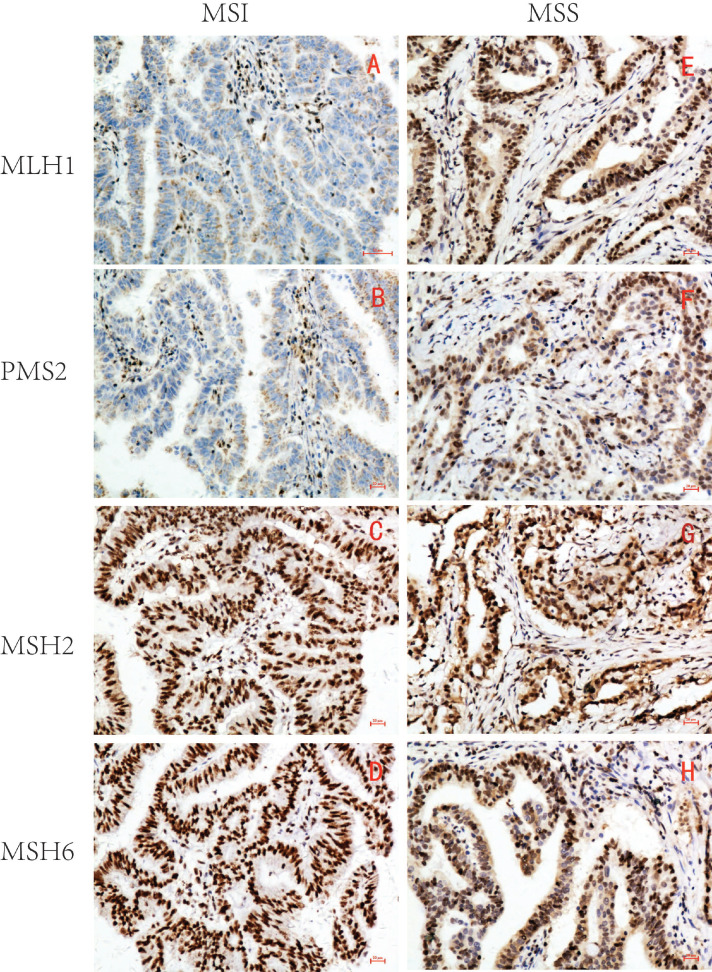
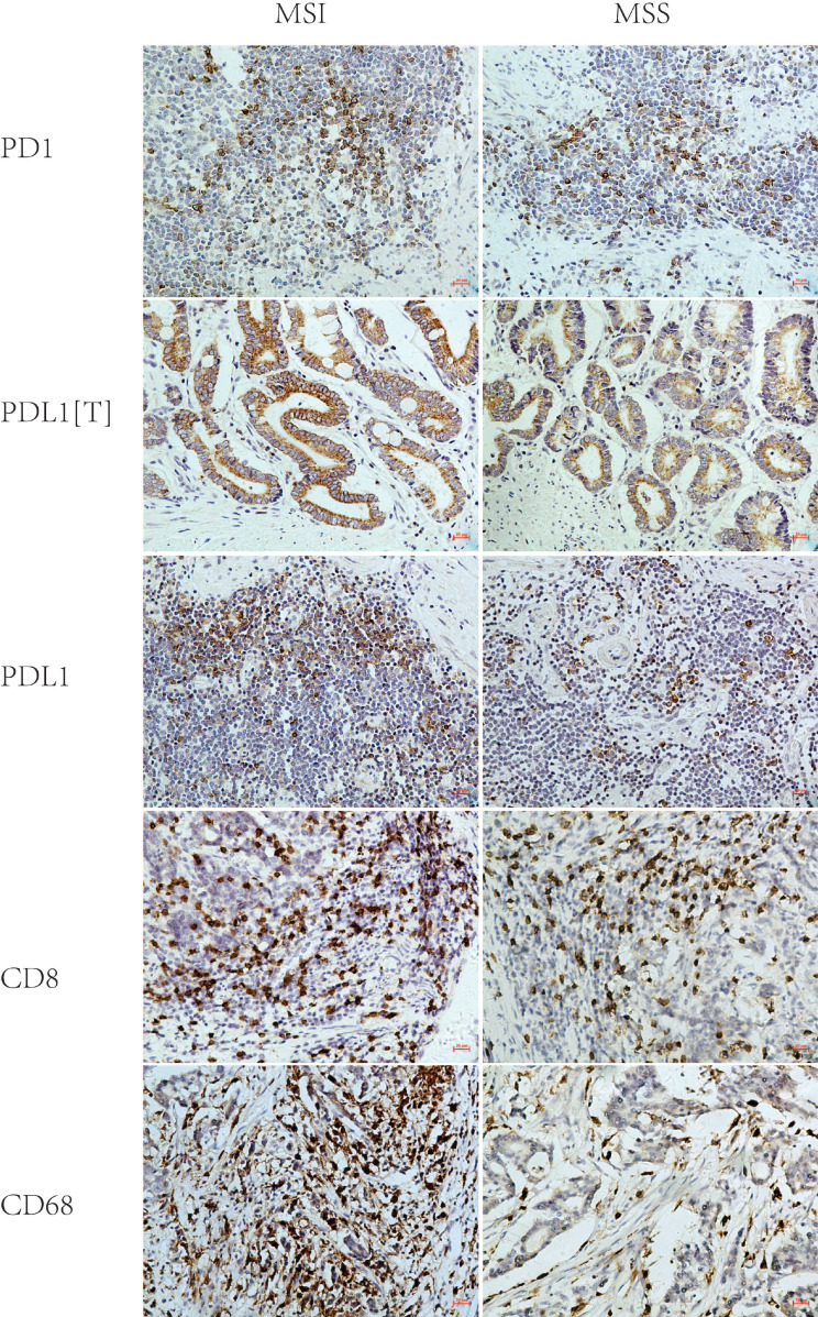
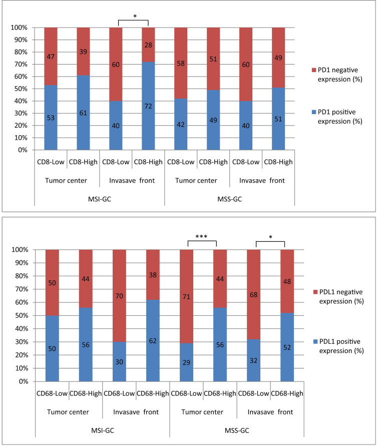
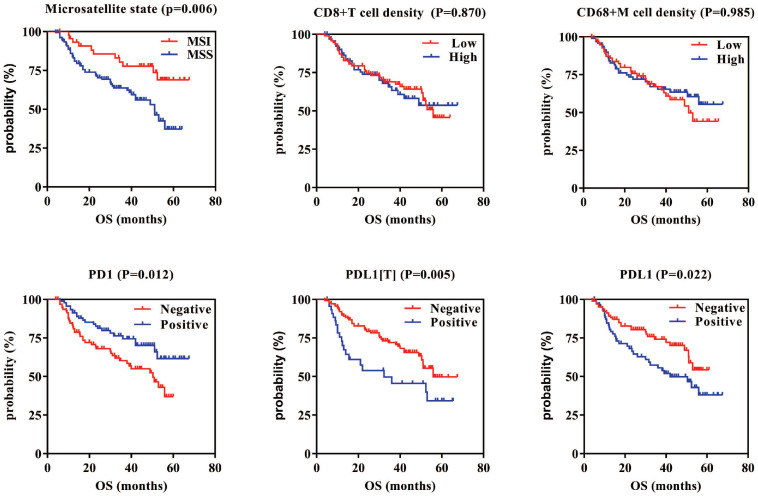
Objective: The microsatellite status and tumor immune microenvironment have a remarkable influence on tumor immunotherapy. This study was performed to investigate programmed cell death protein 1/programmed death ligand 1 (PD1/PDL1) expression and their correlations with CD8+ T cell/CD68+ macrophage (CD68+ M) densities in gastric cancer (GC) at different microsatellite statuses. Methods: The expression of MLH1, PMS2, MSH2, and MSH6 was detected via immunohistochemistry (IHC) to determine the microsatellite status in 215 GC samples obtained from surgical resections. Furthermore, the expression of PD1, PDL1, CD8, and CD68 was detected in the samples via IHC, and the differences and correlations in GC at different microsatellite statuses were then analyzed. PDL1 expression in tumor cells was labeled as PDL1[T], while expression of PD1 and PDL1 in tumor-infiltrating immune cells was labeled as PD1 and PDL1, respectively. Kaplan-Meier analysis was used to evaluate the significance of PD1/PDL1 expression in determining overall survival. Multivariate Cox regression analysis was performed using SPSS software. P-values were determined using the log-rank test. Results: Our results indicated that PD1, PDL1[T], and PDL1 positivity rates were 59%, 35%, and 57% in 46 microsatellite unstable (MSI) GCs and 45%, 22%, and 40% in 169 microsatellite stable (MSS) GCs, respectively. Compared with MSS GC, PD1, PDL1[T], and PDL1 expression was higher in MSI GC (P = 0.109, 0.090, and 0.044, respectively). Additionally, CD8+ T cell and CD68+ M densities were higher in MSI GC than in MSS GC (P = 0.537 and <0.001, respectively). Additionally, CD8+ T cell/CD68+ M densities were evaluated according to tumor center and invasion front. We found that PD1 expression was significantly correlated with CD8+ T cell density at the invasion front of the MSI GC (P = 0.031), whereas PDL1 expression was significantly correlated to high CD68+ M density in the tumor center and invasion front of MSS GC (P = 0.001 and 0.014, respectively). Survival analysis showed that patients with PD1-positive and PDL1[T]/PDL1-negative GC had better prognosis (P = 0.012, 0.005, and 0.022, respectively). Multivariate Cox survival analysis showed that PDL1[T] was an independent prognostic factor for GC. Conclusion: The results suggested that PD1/PDL1 expression and immune response varied at different microsatellite statuses in GC. PD1/PDL1 expression was correlated with CD8+ T cell/CD68+ M densities in GC at different microsatellite statuses, especially at the invasion front. The patients exhibiting high PD1/PDL1 expression or high CD8+ T cell/CD68+ M densities MSI GC might be potential beneficiaries of PD1/PDL1 immunotherapy.
Keywords: CD68; CD8; PD1; PDL1; gastric cancer; tumor immune microenvironment.
© The author(s).
Conflict of interest statement
Competing Interests: The authors have declared that no competing interest exists.
Figures
Similar articles
-
Clinical significance of programmed cell death-ligand 1 expression and the immune microenvironment at the invasive front of colorectal cancers with high microsatellite instability.Int J Cancer. 2018 Feb 15;142(4):822-832. doi: 10.1002/ijc.31107. Epub 2017 Oct 31. Int J Cancer. 2018. PMID: 29044503
-
Analysis of PD1, PDL1, PDL2 expression and T cells infiltration in 1014 gastric cancer patients.Oncoimmunology. 2017 Dec 21;7(3):e1356144. doi: 10.1080/2162402X.2017.1356144. eCollection 2018. Oncoimmunology. 2017. PMID: 29399387 Free PMC article.
-
Prognostic implications of tumor-infiltrating FoxP3+ regulatory T cells and CD8+ cytotoxic T cells in microsatellite-unstable gastric cancers.Hum Pathol. 2014 Feb;45(2):285-93. doi: 10.1016/j.humpath.2013.09.004. Epub 2013 Dec 12. Hum Pathol. 2014. PMID: 24331841
-
The Potential Value of Immunotherapy in Colorectal Cancers: Review of the Evidence for Programmed Death-1 Inhibitor Therapy.Clin Colorectal Cancer. 2016 Dec;15(4):285-291. doi: 10.1016/j.clcc.2016.07.007. Epub 2016 Jul 22. Clin Colorectal Cancer. 2016. PMID: 27553906 Review.
-
PD-1/PD-L1 Checkpoint Inhibitors in Tumor Immunotherapy.Front Pharmacol. 2021 Sep 1;12:731798. doi: 10.3389/fphar.2021.731798. eCollection 2021. Front Pharmacol. 2021. PMID: 34539412 Free PMC article. Review.
Cited by
-
A cohort study using IL-6/Stat3 activity and PD-1/PD-L1 expression to predict five-year survival for patients after gastric cancer resection.PLoS One. 2022 Dec 1;17(12):e0277908. doi: 10.1371/journal.pone.0277908. eCollection 2022. PLoS One. 2022. PMID: 36454780 Free PMC article.
-
Interactions between circRNAs and miR-141 in Cancer: From Pathogenesis to Diagnosis and Therapy.Int J Mol Sci. 2023 Jul 24;24(14):11861. doi: 10.3390/ijms241411861. Int J Mol Sci. 2023. PMID: 37511619 Free PMC article. Review.
-
Mechanism underlying circRNA dysregulation in the TME of digestive system cancer.Front Immunol. 2022 Sep 27;13:951561. doi: 10.3389/fimmu.2022.951561. eCollection 2022. Front Immunol. 2022. PMID: 36238299 Free PMC article. Review.
-
The characteristics and clinical relevance of tumor fusion burden in non-EBV (+) gastric cancer with MSS.BMC Gastroenterol. 2023 May 15;23(1):153. doi: 10.1186/s12876-023-02765-9. BMC Gastroenterol. 2023. PMID: 37189078 Free PMC article.
-
The influence of PD-L1 expression levels on the efficacy of combination therapy in thymic epithelial tumors.Clin Transl Oncol. 2025 Feb;27(2):542-548. doi: 10.1007/s12094-024-03618-x. Epub 2024 Jul 24. Clin Transl Oncol. 2025. PMID: 39046681
References
-
- Bray F, Ferlay J, Soerjomataram I. et al. Global cancer statistics 2018: GLOBOCAN estimates of incidence and mortality worldwide for 36 cancers in 185 countries. CA Cancer J Clin. 2018;68:394–424. - PubMed
LinkOut - more resources
Full Text Sources
Other Literature Sources
Research Materials
Miscellaneous
